. 2009 Mar 27;284(13):8247-55.
doi: 10.1074/jbc.M808492200. Epub 2009 Jan 26.

AMP-activated protein kinase phosphorylates R5/PTG, the glycogen targeting subunit of the R5/PTG-protein phosphatase 1 holoenzyme, and accelerates its down-regulation by the laforin-malin complex

Affiliations

AMP-activated protein kinase phosphorylates R5/PTG, the glycogen targeting subunit of the R5/PTG-protein phosphatase 1 holoenzyme, and accelerates its down-regulation by the laforin-malin complex

Santiago Vernia et al. J Biol Chem. .

Abstract

R5/PTG is one of the glycogen targeting subunits of type 1 protein phosphatase, a master regulator of glycogen synthesis. R5/PTG recruits the phosphatase to the places where glycogen synthesis occurs, allowing the activation of glycogen synthase and the inactivation of glycogen phosphorylase, thus increasing glycogen synthesis and decreasing its degradation. In this report, we show that the activity of R5/PTG is regulated by AMP-activated protein kinase (AMPK). We demonstrate that AMPK interacts physically with R5/PTG and modifies its basal phosphorylation status. We have also mapped the major phosphorylation sites of R5/PTG by mass spectrometry analysis, observing that phosphorylation of Ser-8 and Ser-268 increased upon activation of AMPK. We have recently described that the activity of R5/PTG is down-regulated by the laforin-malin complex, composed of a dual specificity phosphatase (laforin) and an E3-ubiquitin ligase (malin). We now demonstrate that phosphorylation of R5/PTG at Ser-8 by AMPK accelerates its laforin/malin-dependent ubiquitination and subsequent proteasomal degradation, which results in a decrease of its glycogenic activity. Thus, our results define a novel role of AMPK in glycogen homeostasis.


Figures
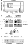
FIGURE 1.
FIGURE 1.
AMPK interacts physically with R5/PTG. A, yeast CTY10.5d cells transformed with plasmids pBTM116 (empty), pBTM-AMPKα2, pBTM-AMPKβ2, and pBTM-AMPKγ1 were co-transformed with plasmids pACT2 (empty), pACT2-GM, pACT2-GL, and pACT2-R5/PTG. Protein interaction was estimated using the yeast two-hybrid system, by measuring the β-galactosidase activity. Values correspond to means from four to six different transformants (bars indicate ± S.D.). B, AMPKα2 co-immunoprecipitates with R5/PTG. Protein extracts (300 μg) were prepared from Hek293 cells transfected with plasmids pCMV-Myc-AMPKα2 and pCMV-HA-R5/PTG. 1 μl of anti-Myc or pre-immune serum (φ) were used to immunoprecipitate the extracts (IP). Pelleted proteins were analyzed by SDS-PAGE and immunodetected with anti-HA (upper panel) and anti-Myc (lower panel) monoclonal antibodies. Proteins in the input crude extracts (CE, 30 μg) were also immunodetected with anti-HA and anti-Myc antibodies. C, interaction of endogenous R5/PTG and AMPKα. Crude extracts (1.5 mg) from mice skeletal muscle (see “Experimental Procedures”) were immunoprecipitated with 1 μl of anti-R5/PTG antibodies or pre-immune serum (φ). Immunoprecipitates were analyzed by SDS-PAGE and Western blotting using anti-AMPKα antibodies. Proteins in the input crude extracts (CE, 50 μg) were also immunodetected with anti-AMPKα and anti-R5/PTG antibodies. D, AMPK phosphorylates a MBP-R5/PTG fusion protein in vitro. MBP-R5/PTG (50 ng) and MBP (100 ng), produced in bacteria were phosphorylated in vitro using 50 milliunits of AMPK (Upstate) and [γ-32P]ATP, in the presence or absence of 300 μm AMP. Samples were analyzed by SDS-PAGE and autoradiography. Size standards are indicated in kilodaltons.
FIGURE 2.
FIGURE 2.
AMPK phosphorylates R5/PTG in vivo. A, cell extracts from CHO cells transfected with plasmid pCMV-HA-R5/PTG were analyzed by two-dimensional electrophoresis and Western blotting using anti-HA monoclonal antibodies (left panel). An aliquot of these extracts was previously treated with λ-phosphatase (middle panel). Cell extracts from CHO cells treated with AICAR (0.5 mm, 6 h) were analyzed similarly (right panel). B, CHO cells were co-transfected with plasmid pCMV-HA-R5/PTG and either a combination of plasmids that reconstituted a constitutively active form of the AMPK complex (pcDNA3-AMPKα2 T172D, pcDNA3-AMPKβ2, and pcDNA3-AMPKγ1; CA-AMPK, right panel) or the same amount of pcDNA3 empty plasmid (left panel). Cell extracts were obtained and analyzed as in A.
FIGURE 3.
FIGURE 3.
AMPK induces the phosphorylation of residues Ser-8 and Ser-268 in R5/PTG. A, 8×His-tagged R5/PTG purified from CHO cells was subjected to mass spectrometry analysis. Ions corresponding to phosphorylated residues are depicted in boldface. B, sequences corresponding to the N and C termini of R5/PTG sequences from different species (GenBank™) were aligned using the ClustalW program. The position of the Ser-8 and Ser-268 is indicated. C, 8×His-tagged R5/PTG was purified from CHO cells grown under conditions of AMPK activation by expressing a constitutively active form of AMPKγ1 (R70Q) subunit. The efficiency of the phosphorylation was compared with that obtained in CHO cells grown under standard conditions (see “Experimental Procedures”). D, cell extracts from CHO cells transfected with plasmid pCMV-HA-R5/PTG S8A S268A were analyzed by two-dimensional electrophoresis and Western blotting using anti-HA monoclonal antibodies.
FIGURE 4.
FIGURE 4.
Phosphorylation status of R5/PTG does not affect its interaction with different partners. Yeast CTY10.5d cells transformed with plasmids pBTM-PP1c, pBTM-AMPKα2, pBTM-AMPKβ2, and pEG202-Laforin were co-transformed with plasmids pACT2 (empty), pACT2-R5/PTG wt, pACT2-R5/PTG S8A, and pACT2-R5/PTG S268A. Protein interaction was estimated using the yeast two-hybrid system, by measuring the β-galactosidase activity. Values correspond to means from four to six different transformants (bars indicate ± S.D.).
FIGURE 5.
FIGURE 5.
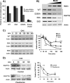
Phosphorylation status of R5/PTG affects its regulation by the laforin/malin complex. A, Hek293 cells were transfected with plasmids expressing the different mutated forms of R5/PTG (pCMV-HA-R5/PTG-derived plasmids) in combination or not with plasmids pCMV-Myc-Laforin and pcDNA-HA-Malin. Twenty-four hours after the transfection, the amount of glycogen was determined as described under “Experimental Procedures.” Bars indicate ± S.D. of three independent experiments. B, degradation profiles of the different mutated forms of R5/PTG. Hek293 cells were co-transfected with pCMV-HA-R5/PTG-derived plasmids, pCMV-Myc-laforin, and different amounts of pcDNA-HA-Malin. Twenty-four hours after the transfection, cell extracts (25 μg) were analyzed by Western blot using anti-HA antibodies; anti-actin was used as loading control; a representative analysis is presented. C, degradation profiles of the different mutated forms of R5/PTG in the presence of cycloheximide. Hek293 cells were co-transfected with pCMV-Myc-laforin, pcDNA3-HA-Malin, and pCMV-HA-R5/PTG plasmids expressing the indicated proteins. Twenty-four hours after the transfection, cycloheximide (CHX, 100 μm) was added to the cultures, and cell extracts were analyzed by Western blotting at different times after the addition, using anti-HA and anti-actin antibodies (left panel); a representative analysis is presented. The relative intensity of the R5/PTG bands respect to the corresponding actin band was plotted as a function of time (right panel). D, degradation profile of R5/PTG in the presence of phenformin. Hek293 cells were co-transfected with pCMV-Myc-laforin, pcDNA3-HA-Malin, and pCMV-HA-R5/PTG plasmids as in B. Twenty-four hours after the transfection, cells were treated with 5 mm phenformin for 2 h, and then cycloheximide (CHX, 100 μm) was added to the cultures. Cell extracts were analyzed by Western blotting at different times after the addition, using anti-HA and anti-actin antibodies (left panel); a representative analysis is presented. The relative intensity of the R5/PTG bands respect to the corresponding actin band was plotted as a function of time (right panel).
FIGURE 6.
FIGURE 6.
Depletion of laforin increases the levels of R5/PTG. A, Hek293 cells were co-transfected with plasmids pCMV-HA-R5/PTG (WT and S8A) and with 1 μg of pSUPER-laf plasmid expressing laforin small interference RNA (see “Experimental Procedures”) or with an empty vector (c-). Twenty-four hours after the transfection, cells extracts (25 μg) were analyzed by Western blotting using anti-HA, anti-laforin, and anti-actin antibodies (right panel). A longer exposure of the blot with anti-HA was used to assess the levels of the proteins in the absence of laforin silencing; a representative analysis is presented. The relative intensity of the R5/PTG bands under control and laforin-depletion conditions in three independent experiments is plotted (left panel); bars indicate ± S.D. B, cell extracts from primary fibroblasts from two LD patients with mutations in the laforin gene (epm2a–/–; Y86X and R241X) and from two healthy subjects (#1 and #2) were analyzed by Western blotting using anti-R5/PTG and anti-laforin and anti-tubulin antibodies (right panel); a representative analysis is presented. The relative intensity of the R5/PTG bands with respect to the corresponding tubulin bands, in three independent experiments, is plotted (left panel); bars indicate ± S.D.
FIGURE 7.
FIGURE 7.
Proposed role of AMPK on glycogen biosynthesis. See text for details. PP1c, catalytic subunit of type 1 protein phosphatase; GS, glycogen synthase; and Ph, glycogen phosphorylase.

Similar articles

Cited by

References

    1. Roach, P. J. (2002) Curr. Mol. Med. 2 101–120 - PubMed
    1. Ferrer, J. C., Favre, C., Gomis, R. R., Fernandez-Novell, J. M., Garcia-Rocha, M., de la Iglesia, N., Cid, E., and Guinovart, J. J. (2003) FEBS Lett. 546 127–132 - PubMed
    1. Chen, Y. H., Hansen, L., Chen, M. X., Bjorbaek, C., Vestergaard, H., Hansen, T., Cohen, P. T., and Pedersen, O. (1994) Diabetes 43 1234–1241 - PubMed
    1. Doherty, M. J., Moorhead, G., Morrice, N., Cohen, P., and Cohen, P. T. (1995) FEBS Lett. 375 294–298 - PubMed
    1. Doherty, M. J., Young, P. R., and Cohen, P. T. (1996) FEBS Lett. 399 339–343 - PubMed

MeSH terms