. 2009 May;182(1):11-23.
doi: 10.1534/genetics.108.098707. Epub 2009 Jan 26.

Tools for fungal proteomics: multifunctional neurospora vectors for gene replacement, protein expression and protein purification

Affiliations

Tools for fungal proteomics: multifunctional neurospora vectors for gene replacement, protein expression and protein purification

Shinji Honda et al. Genetics. 2009 May.

Abstract

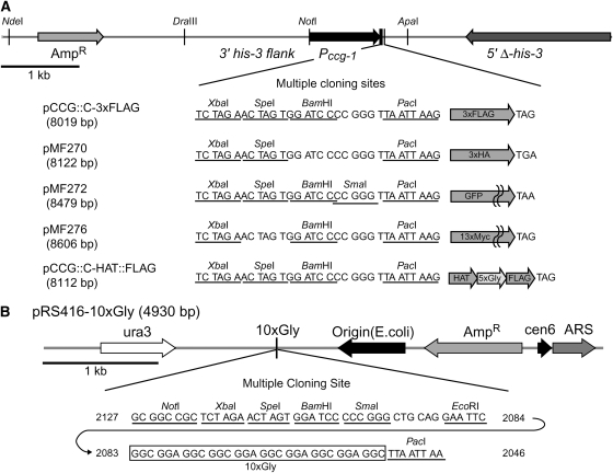
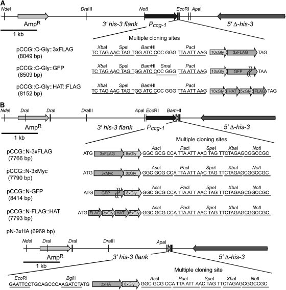
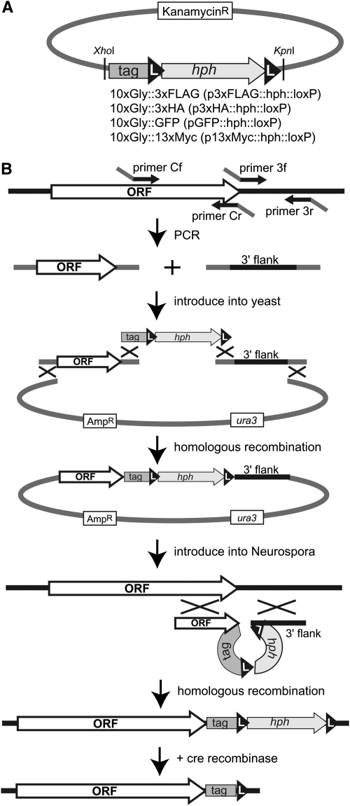
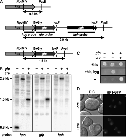
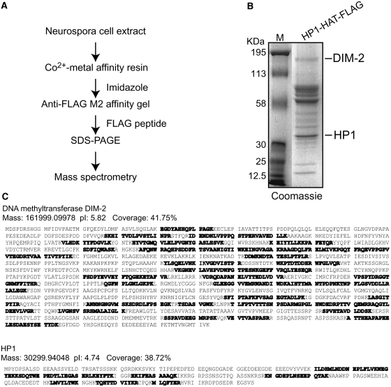
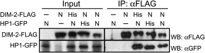
The completion of genome-sequencing projects for a number of fungi set the stage for detailed investigations of proteins. We report the generation of versatile expression vectors for detection and isolation of proteins and protein complexes in the filamentous fungus Neurospora crassa. The vectors, which can be adapted for other fungi, contain C- or N-terminal FLAG, HA, Myc, GFP, or HAT-FLAG epitope tags with a flexible poly-glycine linker and include sequences for targeting to the his-3 locus in Neurospora. To introduce mutations at native loci, we also made a series of knock-in vectors containing epitope tags followed by the selectable marker hph (resulting in hygromycin resistance) flanked by two loxP sites. We adapted the Cre/loxP system for Neurospora, allowing the selectable marker hph to be excised by introduction of Cre recombinase into a strain containing a knock-in cassette. Additionally, a protein purification method was developed on the basis of the HAT-FLAG tandem affinity tag system, which was used to purify HETEROCHROMATIN PROTEIN 1 (HP1) and associated proteins from Neurospora. As expected on the basis of yeast two-hybrid and co-immunoprecipitation (Co-IP) experiments, the Neurospora DNA methyltransferase DIM-2 was found in a complex with HP1. Features of the new vectors allowed for verification of an interaction between HP1 and DIM-2 in vivo by Co-IP assays on proteins expressed either from their native loci or from the his-3 locus.


Figures

F<sc>igure</sc> 1.—
Figure 1.—
Entry and first-generation expression vectors. (A) Partial maps of the expression vectors pCCG∷C-3xFLAG (GenBank accession no. FJ456996), pMF270 (3xHA) (FJ456997), pMF272 (GFP) (AY598428) (Freitag et al. 2004b), pMF276 (13xMyc) (FJ456998), and pCCG∷C-HAT∷FLAG (FJ456999) and sequences of their MCSs. Unique restriction sites are underlined and indicated by vertical bars. This set of vectors contain the N. crassa ccg-1 (Pccg-1) promoter (McNally and Free 1988) and a MCS followed by 3xFLAG, 3xHA, GFP, 13xMyc, or 1xFLAG∷5xGly∷HAT. A gene of interest can be inserted into MCS or inserted with its endogenous promoter between the NotI site and the MCS, replacing Pccg-1. The flanking his-3 sequences allow the construct to be targeted to the N. crassa his-3 locus (Margolin et al. 1997). (B) Map and sequence of the MCS of entry vector pRS416-10xGly (FJ457000). The box marks the 10xGly flexible linker. Genes of interest can be inserted into the MCS using restriction enzymes or by homologous recombination in yeast (Oldenburg et al. 1997). The 8-base cutter PacI site was included to transfer the gene with the poly-glycine linker to epitope tag expression vectors.
F<sc>igure</sc> 2.—
Figure 2.—
Advanced expression vectors with N- or C-terminal epitope tags and a poly-glycine flexible linker. (A) Partial maps of C-terminal epitope tag expression vectors pCCG∷C-Gly∷3xFLAG (FJ457001), pCCG∷C-Gly∷GFP (FJ457002), and pCCG∷C-Gly∷HAT∷FLAG (FJ457003) and sequences of their MCSs. Unique restriction sites are underlined and indicated by vertical bars. All vectors contain the N. crassa ccg-1 promoter (Pccg-1) (McNally and Free 1988), a MCS, and a poly-glycine linker in the same frame and direction. (B) Partial maps of the N-terminal epitope-tag expression vectors pCCG∷N-3xFLAG (FJ457004), pCCG∷N-3xMyc (FJ457005), pCCG∷N-GFP (FJ457006), pCCG∷N-FLAG∷HAT (FJ457007), and pN-3xHA (FJ457008) and sequences of their MCS. Unique restriction sites are underlined and indicated by vertical bars. All vectors except pN-3xHA contain Pccg-1, and pN-3xHA has ApaI, EcoRI, and BglII (ends compatible with those generated by BamHI) sites for introduction of the Pccg-1 or the promoter of a gene of interest. All vectors contain a poly-glycine linker and a MCS in the same frame and direction.
F<sc>igure</sc> 3.—
Figure 3.—
Strategy of creating knock-in constructs containing C-terminal epitope tags and allowing for excision of the selectable marker hph by use of the Cre/loxP system in Neurospora. The knock-in procedure was modified from a gene knock-out procedure described by Colot et al. (2006). (A) Partial map of p3xFLAG∷hphloxP (FJ457009), p3xHA∷ hphloxP (FJ457010), pGFP∷ hphloxP (FJ457011), and p13xMyc∷ hphloxP (FJ457012) plasmids. The epitope tag∷ hphloxP modules, which contain a 10xGly linker and an epitope tag followed by hph (conferring hygR) flanked by loxP (L) sites, is isolated by digestion with XhoI and KpnI. (B) To build custom knock-in constructs, the C-terminal end of the target gene, without the stop codon, and its 3′ flanking region are amplified by PCR from genomic DNA with primers Cf + Cr and 3f + 3r, respectively. Primers Cf and 3r contain 29 nt of identity to the yeast shuttle vector pRS416, and primers Cr and 3f contain 29 nt in common with each end of the knock-in module. The same sequences serve in all the modules so that the same primers (Cf, Cr, 3f, and 3r) work to make knock-in cassettes with any of the epitope tags. The two PCR products, the module, and the linearized pRS416 are cotransformed into yeast to assemble them by homologous recombination (Oldenburg et al. 1997). Circular plasmids containing the knock-in cassette are extracted from the transformants and amplified in E. coli. The knock-in cassette is then isolated and introduced into the Δmus-52 Neurospora strain. Knock-in strains are selected by growth on medium containing hygromycin. Correct integration of the knock-in cassette and expression of the tagged protein in the transformants are confirmed by Southern blotting and Western blotting, respectively. After removing the Δmus-52 marker by crossing, the hph gene flanked by loxP sequences is excised by introduction of linearized pCCG∷Cre (FJ457013), which contains the cre recombinase.
F<sc>igure</sc> 4.—
Figure 4.—
Application of the Cre/loxP system to excise the hph marker in Neurospora. (A) Schematic of the wild-type hpo locus and the hpo locus after integration of the knock-in cassette containing gfp followed by the hph marker flanked by loxP sites (HP1–GFP knock-in cassette) and after excision of the hph marker by introduction of the cre recombinase (see materials and methods). DNA fragments generated by digestion with NgoMIV and PvuII are indicated by arrows, and the regions used as probes for Southern blotting are indicated with horizontal bars. (B) Southern analysis showing integration of the HP1–GFP knock-in cassette at the hpo locus and excision of the hph marker with the Cre/loxP system. Genomic DNA isolated from strains with (+) or without (−) the gfp-tagged hpo and/or in the presence (+) or absence (−) of cre was digested with NgoMIV and PvuII and used for Southern hybridizations with the indicated probes. (C) Sensitivity to hygromycin in strains containing the hpo-gfp knock-in cassette is restored by excision of hph using the Cre/loxP system. Conidia suspensions of strains with (+) or without (−) the gfp-tag construct at hpo and/or in the presence (+) or absence (−) of cre were plated on minimal medium (−) or medium supplemented with histidine (+his) or hygromycin (+hyg) and grown at 32° overnight. (D) Localization of HP1–GFP is not affected by excision of hph using the Cre/loxP system. Conidia of an hpo-gfploxPhphloxP; his-3 strain (−cre, N3322) and an hpo-gfploxP; his-3+Pccg-1cre (+cre, N3684) strain were examined by differential interference contrast (DIC) and fluorescence (HP1–GFP) microscopy.
F<sc>igure</sc> 5.—
Figure 5.—
Purification of HP1-associtated proteins using the HAT–FLAG purification system. (A) Purification and characterization scheme. Cell extracts prepared from strains containing HP1–HAT–FLAG (N3278) were subject to a two-step affinity purification (Co2+ metal affinity purification followed by anti-FLAG affinity purification). (B) HP1 and its associated proteins isolated by the two-step affinity purification system were separated by SDS–PAGE and visualized by Coomassie blue staining. Molecular weights are indicated on the left. DIM-2 and HP1 were identified in excised gel fragments corresponding to bands indicated on the right and subjected to MS analysis. (C) Identification of DIM-2 and HP1 by liquid chromatography-tandem mass spectrometry (LC/MS/MS) analysis. The amino acid sequences, mass, and the isoelectric point (pI) of DIM-2 and HP1 are shown. Peptides detected by LC/MS/MS are indicated in boldface type, and the sequence coverage (percentage of protein sequence detected) is shown.
F<sc>igure</sc> 6.—
Figure 6.—
Co-IP assays of epitope-tagged DIM-2 and HP1 expressed at his-3 or at native loci confirm their interaction. Extracts from strains with or without (−) the FLAG-tagged dim-2 gene and/or the GFP-tagged hpo gene at the his-3 (His) locus or its native loci (N) were immunoprecipitated with anti-FLAG antibodies. Input and immunoprecipitation (IP) samples were fractionated, transferred to PVDF membranes, and immunoblotted with anti-FLAG antibodies or anti-GFP antibodies, as indicated. The tested strains, in order, were N3322, N3323, N3415, N3418, and N3436.

References

    1. Aebersold, R., and M. Mann, 2003. Mass spectrometry-based proteomics. Nature 422 198–207. - PubMed
    1. Bardiya, N., W. G. Alexander, T. D. Perdue, E. G. Barry, R. L. Metzenberg et al., 2008. Characterization of interactions between and among components of the meiotic silencing by unpaired DNA machinery in Neurospora crassa using bimolecular fluorescence complementation. Genetics 178 593–596. - PMC - PubMed
    1. Booher, K. R., and P. Kaiser, 2008. A PCR-based strategy to generate yeast strains expressing endogenous levels of amino-terminal epitope-tagged proteins. Biotech. J. 3 524–529. - PubMed
    1. Borjigin, J., and J. Nathans, 1994. Insertional mutagenesis as a probe of rhodopsin's topography, stability, and activity. J. Biol. Chem. 269 14715–14722. - PubMed
    1. Borkovich, K. A., L. A. Alex, O. Yarden, M. Freitag, G. E. Turner et al., 2004. Lessons from the genome sequence of Neurospora crassa: tracing the path from genomic blueprint to multicellular organism. Microbiol. Mol. Biol. Rev. 68 1–108. - PMC - PubMed

Publication types

MeSH terms