. 2009;3(1):e369.
doi: 10.1371/journal.pntd.0000369. Epub 2009 Jan 27.

The importance of pH in regulating the function of the Fasciola hepatica cathepsin L1 cysteine protease

Affiliations

The importance of pH in regulating the function of the Fasciola hepatica cathepsin L1 cysteine protease

Jonathan Lowther et al. PLoS Negl Trop Dis. 2009.

Abstract

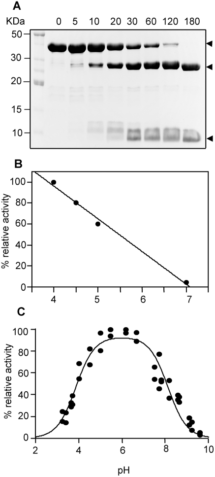
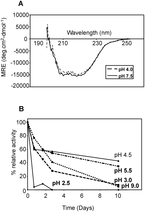
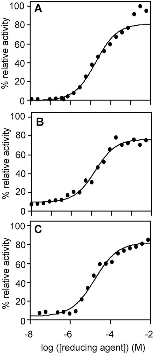
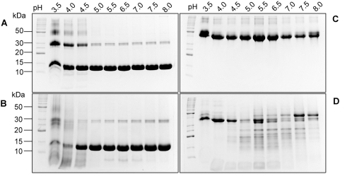
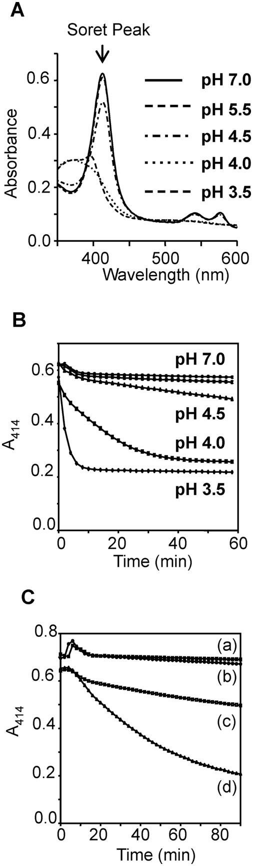
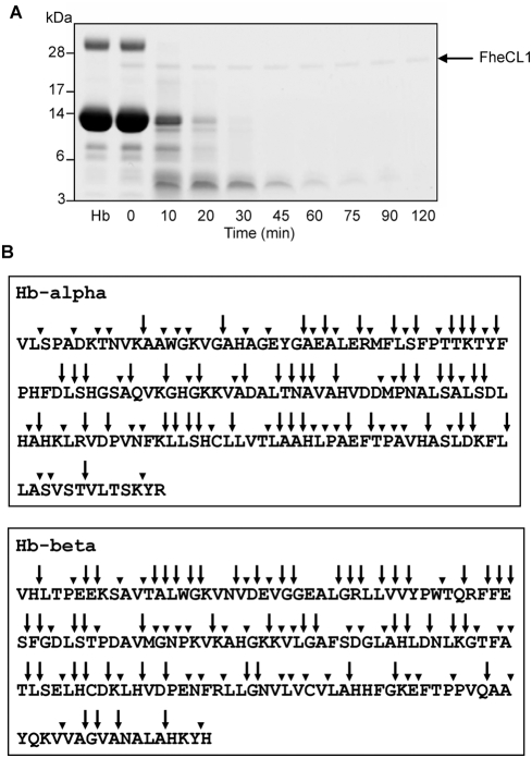
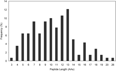
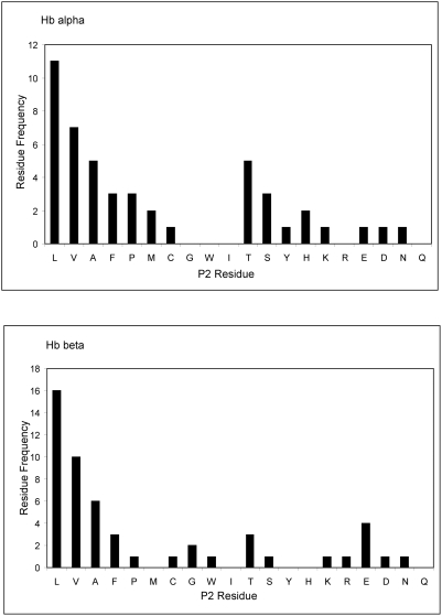
The helminth parasite Fasciola hepatica secretes cathepsin L cysteine proteases to invade its host, migrate through tissues and digest haemoglobin, its main source of amino acids. Here we investigated the importance of pH in regulating the activity and functions of the major cathepsin L protease FheCL1. The slightly acidic pH of the parasite gut facilitates the auto-catalytic activation of FheCL1 from its inactive proFheCL1 zymogen; this process was approximately 40-fold faster at pH 4.5 than at pH 7.0. Active mature FheCL1 is very stable at acidic and neutral conditions (the enzyme retained approximately 45% activity when incubated at 37 degrees C and pH 4.5 for 10 days) and displayed a broad pH range for activity peptide substrates and the protein ovalbumin, peaking between pH 5.5 and pH 7.0. This pH profile likely reflects the need for FheCL1 to function both in the parasite gut and in the host tissues. FheCL1, however, could not cleave its natural substrate Hb in the pH range pH 5.5 and pH 7.0; digestion occurred only at pH</=4.5, which coincided with pH-induced dissociation of the Hb tetramer. Our studies indicate that the acidic pH of the parasite relaxes the Hb structure, making it susceptible to proteolysis by FheCL1. This process is enhanced by glutathione (GSH), the main reducing agent contained in red blood cells. Using mass spectrometry, we show that FheCL1 can degrade Hb to small peptides, predominantly of 4-14 residues, but cannot release free amino acids. Therefore, we suggest that Hb degradation is not completed in the gut lumen but that the resulting peptides are absorbed by the gut epithelial cells for further processing by intracellular di- and amino-peptidases to free amino acids that are distributed through the parasite tissue for protein anabolism.


Conflict of interest statement

The authors have declared that no competing interests exist.

Figures

Figure 1
Figure 1. Influence of pH on zymogen proFheCL1 autocatalytic activation and activity of mature FheCL1.
(A) Analysis by SDS-PAGE of the activation of 0.2 mg/ml FheproCL1 to mature FheCL1 in 0.1 M sodium acetate buffer, pH 4.5. The zymogen, mature enzyme and degraded prosegment are indicated by arrowheads. (B) Kinetic study of the activation of 5 nM FheproCL1 between pH 4.0 and pH 7.0 in the presence of 2 µM Z-Phe-Arg-NHMec. (C) Relative kcat/Km values for the hydrolysis of 0.5 µM Z-Phe-ArgN-Mec by 0.14 nM mature FheCL1 at 37°C.
Figure 2
Figure 2. Stability of the zymogen proFheCL1Gly25 and mature FheCL1 at various pH values.
(A) Far-UV CD spectra of 5.3 µM FheproCL1Gly25 in 50 mM sodium acetate buffer, pH 4.0 and in 50 mM sodium phosphate buffer, pH 7.5. (B) Enzymatic stability of 6.0 µM mature FheCL1 at 37°C and in 0.1 M buffers over the pH range 2.5–9.0. Enzyme activity was monitored at various time-points by diluting aliquots of the reactions into 0.1 M sodium acetate buffer, pH 5.5, containing 3 mM DTT before addition of 5 µM Z-Phe-Arg-NMec.
Figure 3
Figure 3. Effect of small molecular thiols on the activity of FheCL1.
FheCL1 was incubated with (A) DTT, (B) GSH and (C) l-cysteine before adding the fluorogenic substrate Z-Phe-Arg-NHMec. Final assays contained 4 nM enzyme, (10 nM–10 mM) reducing agent and 5 µM substrate in 0.1 M sodium acetate buffer, pH 4.5, with 1 mM EDTA.
Figure 4
Figure 4. pH dependency of FheCL1 hydrolytic activity against protein substrates Hb and ovalbumin.
(A) Hb incubated alone in solutions buffered in the ranges pH 3.5–pH 8.0; (B) Hb incubated with FheCL1 in the same buffers at pH 3.5–pH 8.0; (C) Ovalbumin incubated alone in solutions buffered in the ranges pH 3.5–pH 8.0, and (D) Ovalbumin incubated with FheCL1 in the same buffers at pH 3.5–pH 8.0. Digests were analysed by 15% SDS-PAGE. Molecular size markers are indicated on the left.
Figure 5
Figure 5. Regulation of FheCL1 hydrolytic activity against haemoglobin by pH.
(A) Spectra of 5.0 µM Hb following 1 hr incubation in 0.1 M buffer at pH 3.5, pH 4.0, pH 5.5 and pH 7.0. Decreases in the Soret peak absorbance at 414 nm shows Hb denaturation with decreasing pH. (B) Progress of denaturation of 5.0 µM haemoglobin at several pH values over time as revealed by the decrease in absorbance at 414 nm. (C) Susceptibility of Hb to FheCL1 hydrolytic activity at pH 4.0 and pH 7.0 in the presence 1 mM GSH. (a) 5.0 µM Hb and 1 mM GSH at pH 7.0 (b) 5.0 µM Hb, 1 mM GSH and 1 µM FheCL1 at pH 7.0 (c) 5.0 µM Hb and 1 mM GSH at pH 4.5 (d) 5.0 µM Hb, 1 mM GSH and 1 µM FheCL1 at pH 4.5.
Figure 6
Figure 6. Characterisation of hydrolytic activity of FheCL1 on Hb.
(A) Progress of digestion of Hb by recombinant FheCL1. Purified haemoglobin (Hb, lane 1) was digested by FheCL1 in 0.1 M sodium acetate buffer, pH 4.0, containing 1 mM GSH and 1 mM EDTA at 37°C. Reactions were stopped at time 0 and at various time-points (indicated on x axis) by the addition of the cysteine protease inhibitor E-64 and analysed on 4–12% Bis-Tris NuPage gels. The arrow indicates the position of FheCL1 (25 kDa) that was not degraded in the reaction. Molecular mass markers are shown on the left. (B) Map of Hb α- and β-chains indicating sites of FheCL1 cleavage the substrates. Cleavage sites within Hb present in 10 min reactions (arrows) compared to cleavages that occur with longer incubation times (120 min, arrowheads) as determined by nanoLc-MS/MS.
Figure 7
Figure 7. Analysis of peptides released from Hb following digestion by FheCL1.
Frequency (expressed as a percentage) of peptides of varying length released following proteolysis of Hb alpha and beta chains by FheCL1.
Figure 8
Figure 8. P2 residues in peptides released from Hb following digestion by FheCL1.
Analysis of frequency by which amino acids occur at the P2 position from the peptide bonds cleaved by FheCL1 in Hb α- and β-chains (corresponding to the 10 min reactions shown in Figure 6). The y axis represents the frequency of a particular residue at the P2 position of the haemoglobin substrates and the x axis shows the amino acids as represented by the one-letter code.

References

    1. Andrews SJ. In: Fasciolosis. Dalton JP, editor. Oxford: CABI; 1999. pp. 1–29.
    1. Mas-Coma S, Bargues MD, Valero MA. Fascioliasis and other plant-borne trematode zoonoses. Int J Parasitol. 2005;35:1255–1278. - PubMed
    1. MacManus DP, Dalton JD. Vaccines against the zoonotic trematodes Schistosoma japonicum, Fasciola hepatica and Fasciola gigantica. Parasitology. 2006;133:S43–61. - PubMed
    1. McGonigle L, Mousley A, Marks NJ, Brennan GP, Dalton JP, et al. The silencing of cysteine proteases in Fasciola hepatica newly excysted juveniles using RNA interference reduces gut penetration. Int J Parasitol. 2008;38:149–155. - PubMed
    1. Mulcahy G, O'Connor F, Clery D, Hogan SF, Dowd AJ, et al. Immune responses of cattle to experimental anti-Fasciola hepatica vaccines. Res Vet Sci. 1999;67:27–33. - PubMed

Publication types