. 2008;6(4):636-59.
doi: 10.3390/md6040636. Epub 2008 Dec 22.

Immunomodulatory effects of domoic acid differ between in vivo and in vitro exposure in mice

Affiliations

Immunomodulatory effects of domoic acid differ between in vivo and in vitro exposure in mice

Milton Levin et al. Mar Drugs. 2008.

Abstract

The immunotoxic potential of domoic acid (DA), a well-characterized neurotoxin, has not been fully investigated. Phagocytosis and lymphocyte proliferation were evaluated following in vitro and in vivo exposure to assay direct vs indirect effects. Mice were injected intraperitoneally with a single dose of DA (2.5 microg/g b.w.) and sampled after 12, 24, or 48 hr. In a separate experiment, leukocytes and splenocytes were exposed in vitro to 0, 1, 10, or 100 microM DA. In vivo exposure resulted in a significant increase in monocyte phagocytosis (12-hr), a significant decrease in neutrophil phagocytosis (24-hr), a significant decrease in monocyte phagocytosis (48-hr), and a significant reduction in T-cell mitogen-induced lymphocyte proliferation (24-hr). In vitro exposure significantly reduced neutrophil and monocyte phagocytosis at 1 muM. B- and T-cell mitogen-induced lymphocyte proliferation were both significantly increased at 1 and 10 microM, and significantly decreased at 100 microM. Differences between in vitro and in vivo results suggest that DA may exert its immunotoxic effects both directly and indirectly. Modulation of cytosolic calcium suggests that DA exerts its effects through ionotropic glutamate subtype surface receptors at least on monocytes. This study is the first to identify DA as an immunotoxic chemical in a mammalian species.

Keywords: Domoic acid; adaptive immunity; immunotoxicity; innate immunity.


Figures

Figure 1.
Figure 1.
Flow cytometric dot plot of mouse peripheral blood leukocytes. Sub-populations of leukocytes can be easily distinguished based on forward scatter (relative size) and side scatter (relative complexity). Neutrophils (R1) are large and complex; lymphocytes (R3) are smaller and less complex, while monocytes (R2) are slightly larger than lymphocytes and less complex than neutrophils.
Figure 2.
Figure 2.
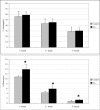
In vivo peripheral blood neutrophil (top) and monocyte (bottom) phagocytosis (mean +SD) in unexposed (control; n=10) and DA-exposed mice (n=10) 12 hr after exposure (t-test: *, p < 0.05).
Figure 3.
Figure 3.
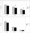
In vivo peripheral blood neutrophil (top) and monocyte (bottom) phagocytosis (mean +SD) in unexposed (control; n=10) and DA-exposed mice (n=10) 24 hr after exposure (t-test: *, p < 0.05).
Figure 4.
Figure 4.
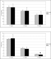
In vivo peripheral blood neutrophil (top) and monocyte (bottom) phagocytosis (mean +SD) in unexposed (control; n=10) and DA-exposed mice (n=10) 48 hr after exposure (t-test: *, p < 0.05).
Figure 5.
Figure 5.
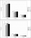
In vivo mitogen-induced lymphocyte (splenocyte) proliferation (mean +SD) in unexposed (control; n=10) and DA-exposed mice (n=10) 24 hr after exposure (t-test, *p < 0.05).
Figure 6.
Figure 6.
In vitro peripheral blood neutrophil (top) and monocyte (bottom) phagocytosis (mean +SD) with increasing concentrations of DA (n=10; RM ANOVA, *p < 0.05).
Figure 7.
Figure 7.
In vitro mitogen-induced lymphocyte (splenocyte) proliferation (mean +SD) with increasing concentrations of DA (n=10; RM ANOVA, *p < 0.05).
Figure 8.
Figure 8.
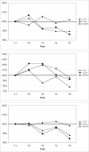
Changes in peripheral blood neutrophil (top), monocyte (middle), and lymphocyte (bottom) cytosolic calcium upon exposure to increasing concentrations of domoic acid. Data are expressed as the % of the unexposed control over time (T-1: 1 min prior to exposure, T0: time of exposure to agonists, T2: 2 min after exposure, T4: 4 min after exposure, T6: 6 min after exposure). For each time point, data are presented as the average of 3 mice. (100% indicated by dotted line)
Figure 9.
Figure 9.
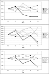
Changes in peripheral blood neutrophil (top), monocyte (middle), and lymphocyte (bottom) cytosolic calcium upon exposure to the ionotropic glutamate receptor agonists, L-glutamate, kainate, AMPA, NMDA, as well as domoic acid (all at 1 μM). Data are expressed as the % of the unexposed control over time (T-1: 1 min prior to exposure, T0: time of exposure to agonists, T2: 2 min after exposure, T4: 4 min after exposure, T6: 6 min after exposure). For each time point, data are presented as the average of 3 mice per agonists. (100% indicated by dotted line)

References

    1. Van Dolah FM. Marine algal toxins: origins, health effects, and their increased occurrence. Environ. Health Perspect. 2000;108(Suppl 1):133–141. - PMC - PubMed
    1. Sayer A, Hu Q, Bourdelais AJ, Baden DG, Gibson JE. The effect of brevenal on brevetoxin-induced DNA damage in human lymphocytes. Arch. Toxicol. 2005;79(11):683–688. - PMC - PubMed
    1. Shen PP, Zhao SW, Zheng WJ, Hua ZC, Shi Q, Liu ZT. Effects of cyanobacteria bloom extract on some parameters of immune function in mice. Toxicol. Lett. 2003;143(1):27–36. - PubMed
    1. Walsh CJ, Luer CA, Noyes DR . Effects of environmental stressors on lymphocyte proliferation in Florida manatees, Trichechus manatus latirostris. Vet. Immunol. Immunopathol. 2005;103(3–4):247–256. - PubMed
    1. Witkowski JM, Siebert J, Lukaszuk K, Trawicka L. Comparison of effect of a panel of membrane channel blockers on the proliferative, cytotoxic and cytoadherence abilities of human peripheral blood lymphocytes. Immunopharmacol. 1993;26(1):53–63. - PubMed

MeSH terms

LinkOut - more resources