

Fibronectin and focal adhesion kinase small interfering RNA modulate rat retinal Müller cells adhesion and migration
- PMID: 19172391
- PMCID: PMC11505797
- DOI: 10.1007/s10571-009-9346-x
Fibronectin and focal adhesion kinase small interfering RNA modulate rat retinal Müller cells adhesion and migration
Abstract
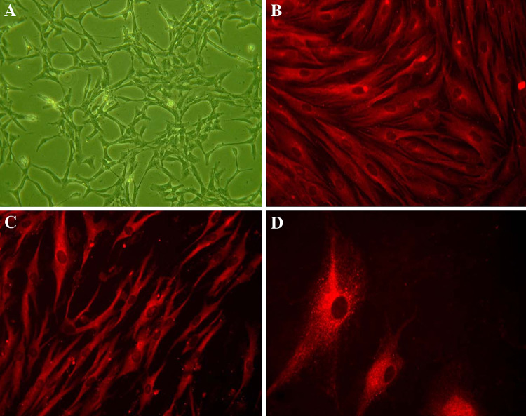
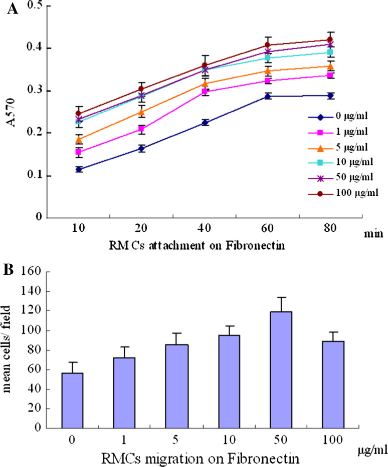
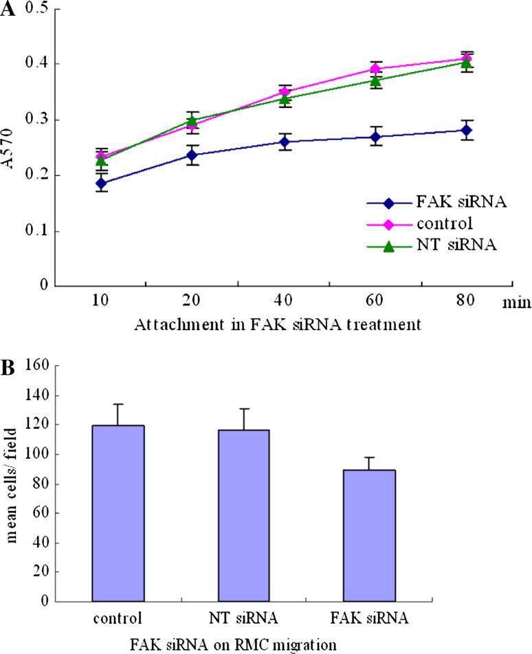
Retinal Müller cells (RMCs) hypertrophy and proliferation play a crucial role in epiretinal membrane formation. This study was designed to analyze the effects of Fibronectin and specific FAK siRNA in cell adhesion and migration in rat Müller cells. RMCs were cultured and identified by GFAP, Vimentin, and GLAST mAb, respectively. The cells were planted on dishes coated with Fibronectin at 0, 1, 5, 10, 50, and 100 microg/ml. The attachment and migration assay was applied to characterize the RMCs-Fibronectin interactions. Cell lysis and Western blotting were utilized to detect beta(1)-integrin, FAK, and GLAST protein expression. Then the cells were treated with FAK siRNA, non-targeting siRNA, and control medium. The cell cycle and apoptosis rate was determined by flow cytometry. The attachment, migration, and Western blotting assay were repeated. These data suggested that almost all the cells expressed GFAP, Vimentin, and GLAST, respectively, which ensured most of the harvested cells were RMCs. In attachment assay, the A570 values increased significantly with time (F = 1105.439, P < 0.001) and Fibronectin concentration (F = 424.683, P < 0.001). There were significant difference between each Fibronectin concentration in RMCs migration (F = 34.703, P < 0.000). The expression ratio of FAK, beta(1)-integrin, and GLAST elevated significantly as Fibronectin concentration increased (F = 54.755, P < 0.000; F = 119.962, P < 0.000; F = 39.287, P < 0.000). The Fibronectin pretreatment was settled on 50 microg/ml for siRNA inhibition assays. The specific FAK siRNA treatment significantly increased G(0)/G(1) percentage and apoptosis rate compared with NT siRNA and control group (F = 11.526, P = 0.009; F = 64.772, P < 0.000). The apoptotic rate was significantly suppressed by inhibitors of caspase-8 and 3 (F = 10.500, P = 0.011). The A570 values were significantly suppressed in FAK siRNA groups compared with NT siRNA and control group (F = 154.241, P < 0.000), and the mean migratory cells per view field were significantly decreased (F = 10.906, P = 0.001). FAK and GLAST expression ratio decreased significantly after FAK siRNA treatment (F = 5.315, P = 0.047; F = 5.985, P = 0.042). Take together, FAK is involved in beta(1)-integrin mediated adhesive signaling and play a critical role in regulating Müller cell adhesion, migration, and so far as to glutamate transportation functions.
Figures
Similar articles
-
Optimal intensity shock wave promotes the adhesion and migration of rat osteoblasts via integrin β1-mediated expression of phosphorylated focal adhesion kinase.J Biol Chem. 2012 Jul 27;287(31):26200-12. doi: 10.1074/jbc.M112.349811. Epub 2012 May 31. J Biol Chem. 2012. PMID: 22654119 Free PMC article.
-
Fibronectin promotes migration and invasion of ovarian cancer cells through up-regulation of FAK-PI3K/Akt pathway.Cell Biol Int. 2014 Jan;38(1):85-91. doi: 10.1002/cbin.10184. Epub 2013 Oct 17. Cell Biol Int. 2014. PMID: 24115647
-
Targeting Pyk2 to beta 1-integrin-containing focal contacts rescues fibronectin-stimulated signaling and haptotactic motility defects of focal adhesion kinase-null cells.J Cell Biol. 2001 Jan 8;152(1):97-110. doi: 10.1083/jcb.152.1.97. J Cell Biol. 2001. PMID: 11149924 Free PMC article.
-
The role of focal adhesion kinase in transforming growth factor-β2 induced migration of human lens epithelial cells.Int J Mol Med. 2018 Dec;42(6):3591-3601. doi: 10.3892/ijmm.2018.3912. Epub 2018 Oct 2. Int J Mol Med. 2018. PMID: 30280182
-
Integrins in cell adhesion and signaling.Hum Cell. 1996 Sep;9(3):181-6. Hum Cell. 1996. PMID: 9183647 Review.
Cited by
-
Immunological Insights in Equine Recurrent Uveitis.Front Immunol. 2021 Jan 8;11:609855. doi: 10.3389/fimmu.2020.609855. eCollection 2020. Front Immunol. 2021. PMID: 33488614 Free PMC article. Review.
-
Osteopontin and fibronectin levels are decreased in vitreous of autoimmune uveitis and retinal expression of both proteins indicates ECM re-modeling.PLoS One. 2011;6(12):e27674. doi: 10.1371/journal.pone.0027674. Epub 2011 Dec 14. PLoS One. 2011. PMID: 22194789 Free PMC article.
-
Osteopontin Regulates AQP4 Expression by TRPV4 Activation in Müller Cells: Implications for Retinal Homeostasis.Mol Neurobiol. 2025 Apr;62(4):4769-4784. doi: 10.1007/s12035-024-04595-6. Epub 2024 Nov 1. Mol Neurobiol. 2025. PMID: 39485629
References
-
- Bringmann A, Reichenbach A (2001) Role of Müller cells in retinal degenerations. Front Biosci 6:E72–E92. doi:10.2741/Bringman - PubMed
-
- Bringmann A, Francke M, Pannicke T, Biedermann B, Kodal H, Faude F, Reichelt W (2006) Müller cells in the healthy and diseased retina. Prog Retin Eye Res 25:397–424. doi:10.1016/j.preteyeres.2006.05.003 - PubMed
-
- Carlson MA, Prall AK, Gums JJ (2007) RNA interference in human foreskin fibroblasts within the three-dimensional collagen matrix. Mol Cell Biochem 306(1–2):123–132. doi:10.1007/s11010-007-9561-z - PubMed
-
- Charteris DG, Sethi CS, Lewis GP, Fisher SK (2002) Proliferative vitreoretinopathy: developments in adjunctive treatment and retinal pathology. Eye 16:369–374. doi:10.1038/sj.eye.6700194 - PubMed
-
- Claudepierre T, Rodius F, Frasson M, Fontaine V, Picaud S, Dreyfus H, Mornet D, Rendon A (1999) Differential distribution of dystrophins in rat retina. Invest Ophthalmol Vis Sci 40:1520–1529 - PubMed
MeSH terms
Substances
LinkOut - more resources
Full Text Sources
Molecular Biology Databases
Research Materials
Miscellaneous
