

Volume and iron content in basal ganglia and thalamus
- PMID: 19172651
- PMCID: PMC6871035
- DOI: 10.1002/hbm.20698
Volume and iron content in basal ganglia and thalamus
Abstract
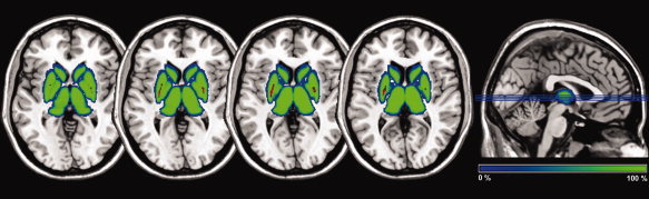
Magnetic resonance imaging (MRI) studies have highlighted the possibility to investigate brain iron content in vivo. In this study, we combined T2* relaxometry and automatic segmentation of basal ganglia based on T1-weighted images in healthy subjects, with the aim of characterizing age related changes in volume and iron-related relaxivity values (R2*) of these structures. Thirty healthy subjects underwent MR imaging at 3 Tesla. Mean R2* values and volumes were calculated for the selected subcortical structures (pallidum, putamen, thalamus and caudate nucleus). Our results showed a correlation between R2* values and iron concentration as calculated from published post-mortem data. Furthermore, we observed a shrinkage/iron increase with a different pattern in the anatomical regions selected in this work, suggesting that the age-related changes on these MR parameters are specific to the subcortical structure considered. In particular, the putamen demonstrated a decrease of volume and an increase of iron level, with the posterior region of this structure appearing more disposed to iron deposition. Our work suggests that combining volumetry and iron estimation in MRI permits to investigate in vivo neurophysiological and neuropathological changes of basal ganglia.
(c) 2009 Wiley-Liss, Inc.
Figures


Comment in
-
Letter to the editor: Brain iron mapping using MRI relaxation rate or R₂* revisited.Hum Brain Mapp. 2012 Aug;33(8):2003-4. doi: 10.1002/hbm.21328. Epub 2011 Aug 5. Hum Brain Mapp. 2012. PMID: 21823205 Free PMC article. No abstract available.
References
-
- Ahsan RL,Allom R,Gousias IS,Habib H,Turkheimer FE,Free S,Lemieux L,Myers R,Duncan JS,Brooks DJ,Koepp MJ,Hammers A ( 2007): Volumes, spatial extents and a probabilistic atlas of the human basal ganglia and thalamus. Neuroimage 38: 261–270. - PubMed
-
- Anastasi G,Cutroneo G,Tomasello F,Lucerna S,Vitetta A,Bramanti P,Di Bella P,Parenti A,Porzionato A,Macchi V, et al. ( 2006): In vivo basal ganglia volumetry through application of NURBS models to MR images. Neuroradiology 48(5): 338–345. - PubMed
-
- Bartzokis G,Beckson M,Hance DB,Marx P,Foster JA,Marder SR ( 1997): MR evaluation of age‐related increase of brain iron in young adult and older normal males. Magn Reson Imaging 15(1): 29–35. - PubMed
-
- Bartzokis G,Cummings J,Perlman S,Hance DB,Mintz J ( 1999a): Increased basal ganglia iron levels in Huntington disease. Arch Neurol 56(5): 569–574. - PubMed
-
- Bartzokis G,Cummings JL,Markham CH,Marmarelis PZ,Treciokas LJ,Tishler TA,Marder SR,Mintz J ( 1999b): MRI evaluation of brain iron in earlier‐ and later‐onset Parkinson's disease and normal subjects. Magn Reson Imaging 17(2): 213–222. - PubMed
Publication types
MeSH terms
Substances
LinkOut - more resources
Full Text Sources
Other Literature Sources
Medical
