

Review
doi: 10.1038/nri2381.
Formation and function of the lytic NK-cell immunological synapse
Affiliations
- PMID: 19172692
- PMCID: PMC2772177
- DOI: 10.1038/nri2381
Item in Clipboard
Review
Formation and function of the lytic NK-cell immunological synapse
Nat Rev Immunol.
2008 Sep.
Abstract
The natural killer (NK)-cell immunological synapse is the dynamic interface formed between an NK cell and its target cell. Formation of the NK-cell immunological synapse involves several distinct stages, from the initiation of contact with a target cell to the directed delivery of lytic-granule contents for target-cell lysis. Progression through the individual stages is regulated, and this tight regulation underlies the precision with which NK cells select and kill susceptible target cells (including virally infected cells and cancerous cells) that they encounter during their routine surveillance of the body.
Figures
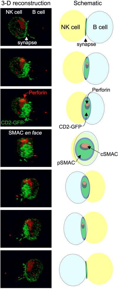
The mature natural killer (NK)-cell lytic synapse is defined by the formation of a supramolecular activation cluster (SMAC) at the interface between the NK-cell and target cell to which lytic granules polarize. The prototypical version of this synapse contains a central SMAC (cSMAC) that includes a secretory domain through which lytic granules may traverse. The series of images show a YTS human NK cell expressing a CD2-GFP (green fluorescent protein) fusion protein making contact with an EBV-transfomed B-cell line. The cell-cell conjugates were fixed, permeabilized and evaluated for the presence of perforin (red) using a monoclonal antibody. Perforin is contained within lytic granues and is presented as a surrogate for them, while CD2 patterning under normal conditions parallels F-actin at the mature synapse. Serial images were obtained using a confocal microscope through the z-axis and the three-dimensional cell volume reconstructed in silico. The individual images on the left from top to bottom demonstrate a 180-degree rotation around the z-axis (Scale bar=5μM) The schematic on the right displays the position of the NK cell (yellow) and B cell (blue), CD2 at the SMAC (green) and perforin at the SMAC (red) in each image. Also see an interactive version in online Supplementary Figure 1.

It is proposed that the formation of a functional natural killer (NK)-cell lytic synapse can be divided into three main stages — recognition, effector and termination stages — that are each subdivided into multiple steps. Important steps proposed to occur in the recognition stage include adhesion and initial activation signaling. In the effector stage key steps include actin reorganization, receptor clustering, MTOC and lytic granule polarization, and lytic granule fusion with the plasma membrane. In the termination stage crucial steps are proposed to include a period of inactivity and detachment. The specific time required to progress through the various stages varies and is likely to be a feature of the given target cell as well as the activation state of the NK cell. The linearity of connections between certain steps is supported by experimental data as outlined in the text, such as a requirement for actin reorganization for MTOC polarization. In others, however, linearity is inferred and is presented as a proposed model. The inhibitory synapse (Box 3) has been shown to halt progression of the lytic synapse by interfering with the late recognition stage and early effector stage – specifically activation signaling, actin reorganization and receptor clustering steps.
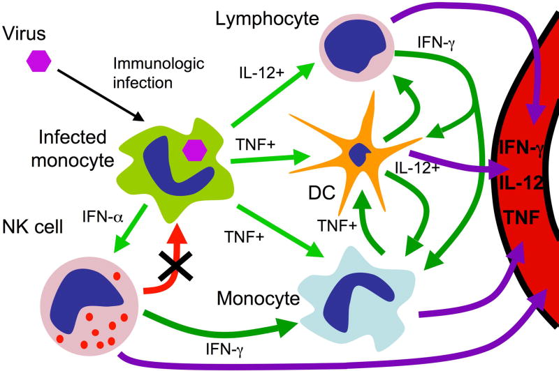
Pathogenic viral infection in a normal individual leads to the production of pro-inflammatory factors, such as tumour-necrosis factor (TNF), interferon-α (IFNα) and interleukin-12 (IL-12) by the infected cell (light green arrows). This induces relevant responses from uninfected cells including dendritic cells, monocytes, natural killer (NK) cells and other lymphocytes. These cells produce additional factors (dark green arrows) to further activate the induced cells and elicit the responses of others. Once induced, NK-cell cytotoxicity can help to rapidly eliminate the infected cell (thick red arrow) and serves to prevent further immune-cell activation induced by the infected cell. NK cell cytotoxicity can also eliminate other non-infected, activated monocytes and dendritic cells to provide additional and critical immunoregulatory function (thin red arrows). These processes have the potential to remain localized and focused to sites of infection. In an individual with impaired NK-cell cytotoxicity but a normal capacity for NK-cell activation, the NK-cell lytic synapse directed against the infected cell does not eliminate it (x). So, the inflammatory response continues unabated, leads to further activation of uninfected cells, and further pro-inflammatory activity from the induced cells. Although the infection may be localized, and the initial responding cells localized to the infection, the response amplifies and leads to an uncontrolled systemic inflammatory syndrome (purple arrows). The inflammatory response may control the viral replication, but without eliminating the source the inflammation itself may not be containable.
References
-
- Orange JS, Ballas ZK. Natural killer cells in human health and disease. Clin Immunol. 2006;118:1–10. - PubMed
-
- Wulfing C, Purtic B, Klem J, Schatzle JD. Stepwise cytoskeletal polarization as a series of checkpoints in innate but not adaptive cytolytic killing. Proc Natl Acad Sci U S A. 2003;100:7767–72. Introduction of steps in NK cell immunological synapse formation centered around the cytoskeleton (see also #12) with critical comparisons to T cells underscoring differences. - PMC - PubMed
-
- Grakoui A, et al. The immunological synapse: a molecular machine controlling T cell activation. Science. 1999;285:221–7. - PubMed
-
- Monks CR, Freiberg BA, Kupfer H, Sciaky N, Kupfer A. Three-dimensional segregation of supramolecular activation clusters in T cells. Nature. 1998;395:82–6. - PubMed
-
- Davis DM, Dustin ML. What is the importance of the immunological synapse? Trends Immunol. 2004;25:323–7. - PubMed
Publication types
MeSH terms
Grants and funding
LinkOut - more resources
Full Text Sources
Other Literature Sources
