

myo-Inositol oxygenase: a radical new pathway for O(2) and C-H activation at a nonheme diiron cluster
- PMID: 19173070
- PMCID: PMC2788986
- DOI: 10.1039/b811885j
myo-Inositol oxygenase: a radical new pathway for O(2) and C-H activation at a nonheme diiron cluster
Abstract
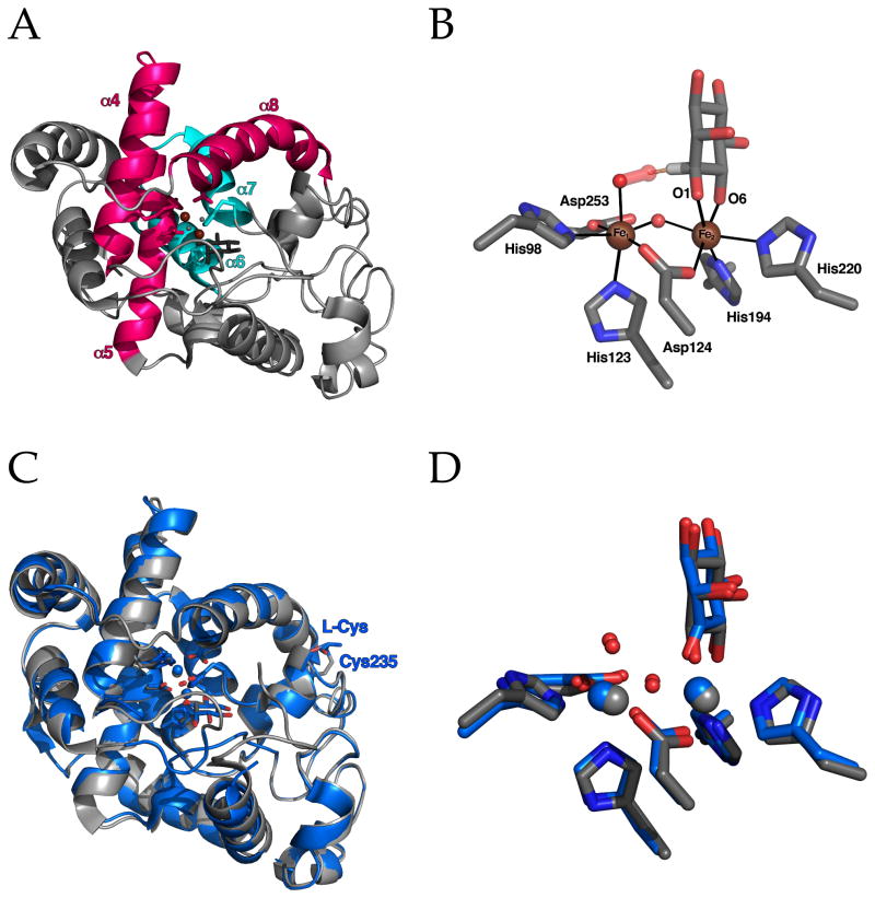
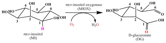
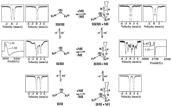
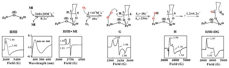
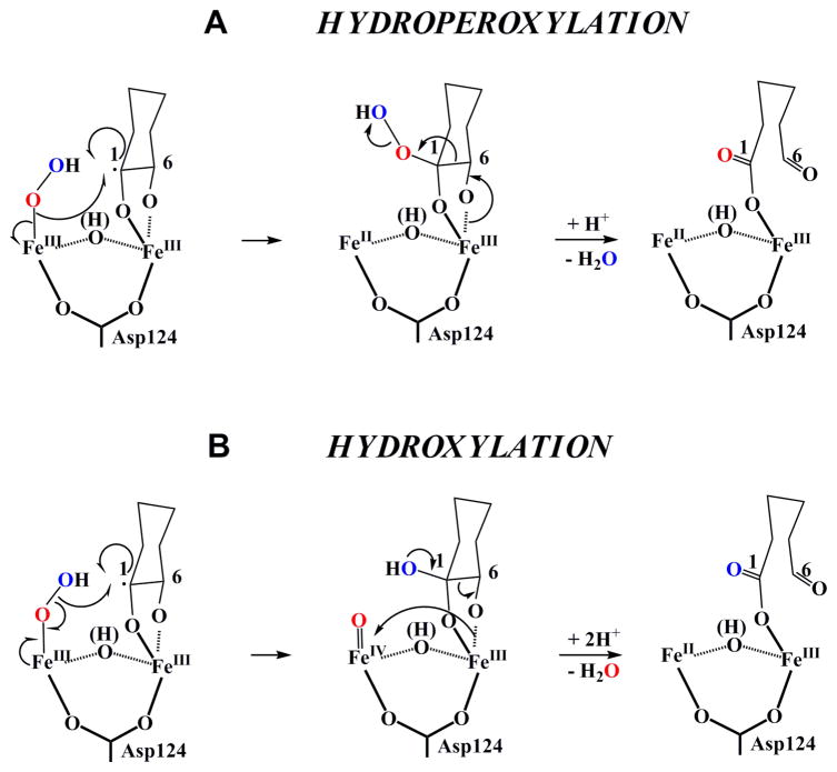
The enzyme myo-inositol oxygenase (MIOX) catalyzes conversion of myo-inositol (cyclohexan-1,2,3,5/4,6-hexa-ol or MI) to d-glucuronate (DG), initiating the only known pathway in humans for catabolism of the carbon skeleton of cell-signaling inositol (poly)phosphates and phosphoinositides. Recent kinetic, spectroscopic and crystallographic studies have shown that the enzyme activates its substrates, MI and O(2), at a carboxylate-bridged nonheme diiron(ii/iii) cluster, making it the first of many known nonheme diiron oxygenases to employ the mixed-valent form of its cofactor. Evidence suggests that: (1) the Fe(iii) site coordinates MI via its C1 and C6 hydroxyl groups; (2) the Fe(ii) site reversibly coordinates O(2) to produce a superoxo-diiron(iii/iii) intermediate; and (3) the pendant oxygen atom of the superoxide ligand abstracts hydrogen from C1 to initiate the unique C-C-bond-cleaving, four-electron oxidation reaction. This review recounts the studies leading to the recognition of the novel cofactor requirement and catalytic mechanism of MIOX and forecasts how remaining gaps in our understanding might be filled by additional experiments.
Figures
Similar articles
-
Oxygen activation by a mixed-valent, diiron(II/III) cluster in the glycol cleavage reaction catalyzed by myo-inositol oxygenase.Biochemistry. 2006 May 2;45(17):5402-12. doi: 10.1021/bi0526276. Biochemistry. 2006. PMID: 16634621
-
Evidence for C-H cleavage by an iron-superoxide complex in the glycol cleavage reaction catalyzed by myo-inositol oxygenase.Proc Natl Acad Sci U S A. 2006 Apr 18;103(16):6130-5. doi: 10.1073/pnas.0508473103. Epub 2006 Apr 10. Proc Natl Acad Sci U S A. 2006. PMID: 16606846 Free PMC article.
-
Demonstration by 2H ENDOR spectroscopy that myo-inositol binds via an alkoxide bridge to the mixed-valent diiron center of myo-inositol oxygenase.J Am Chem Soc. 2006 Aug 16;128(32):10374-5. doi: 10.1021/ja063602c. J Am Chem Soc. 2006. PMID: 16895396
-
Hydroxylation of C-H bonds at carboxylate-bridged diiron centres.Philos Trans A Math Phys Eng Sci. 2005 Apr 15;363(1829):861-77; discussion 1035-40. doi: 10.1098/rsta.2004.1532. Philos Trans A Math Phys Eng Sci. 2005. PMID: 15901540 Review.
-
Potential of engineering the myo-inositol oxidation pathway to increase stress resilience in plants.Mol Biol Rep. 2022 Aug;49(8):8025-8035. doi: 10.1007/s11033-022-07333-0. Epub 2022 Mar 16. Mol Biol Rep. 2022. PMID: 35294703 Review.
Cited by
-
Factors affecting the carboxylate shift upon formation of nonheme diiron-O2 adducts.Inorg Chem. 2013 Mar 4;52(5):2627-36. doi: 10.1021/ic302543n. Epub 2013 Feb 22. Inorg Chem. 2013. PMID: 23432330 Free PMC article.
-
Anaerobic functionalization of unactivated C-H bonds.Curr Opin Chem Biol. 2009 Feb;13(1):58-73. doi: 10.1016/j.cbpa.2009.02.036. Epub 2009 Mar 16. Curr Opin Chem Biol. 2009. PMID: 19297239 Free PMC article. Review.
-
Formation of Unstable and very Reactive Chemical Species Catalyzed by Metalloenzymes: A Mechanistic Overview.Molecules. 2019 Jul 4;24(13):2462. doi: 10.3390/molecules24132462. Molecules. 2019. PMID: 31277490 Free PMC article. Review.
-
Biosynthesis of fosfomycin in pseudomonads reveals an unexpected enzymatic activity in the metallohydrolase superfamily.Proc Natl Acad Sci U S A. 2021 Jun 8;118(23):e2019863118. doi: 10.1073/pnas.2019863118. Proc Natl Acad Sci U S A. 2021. PMID: 34074759 Free PMC article.
-
The Microbial Degradation of Natural and Anthropogenic Phosphonates.Molecules. 2023 Sep 29;28(19):6863. doi: 10.3390/molecules28196863. Molecules. 2023. PMID: 37836707 Free PMC article. Review.
References
-
- Wallar BJ, Lipscomb JD. Chem Rev. 1996;96:2625–2657. - PubMed
-
- Solomon EI, Brunold TC, Davis MI, Kemsley JN, Lee SK, Lehnert N, Neese F, Skulan AJ, Yang YS, Zhou J. Chem Rev. 2000;100:235–349. - PubMed
-
- Merkx M, Kopp DA, Sazinsky MH, Blazyk JL, Müller J, Lippard SJ. Angew Chem, Int Ed Engl. 2001;40:2782–2807. - PubMed
-
- Fox BG, Lyle KS, Rogge CE. Acc Chem Res. 2004;37:421–429. - PubMed
-
- Krebs C, Price JC, Baldwin J, Saleh L, Green MT, Bollinger JM., Jr Inorg Chem. 2005;44:742–757. - PubMed
Publication types
MeSH terms
Substances
Grants and funding
LinkOut - more resources
Full Text Sources
Medical