. 2009 Feb 24;48(7):1462-73.
doi: 10.1021/bi801771g.

Essential role of c-Cbl in amphiregulin-induced recycling and signaling of the endogenous epidermal growth factor receptor

Affiliations

Essential role of c-Cbl in amphiregulin-induced recycling and signaling of the endogenous epidermal growth factor receptor

Aleksander Baldys et al. Biochemistry. .

Abstract

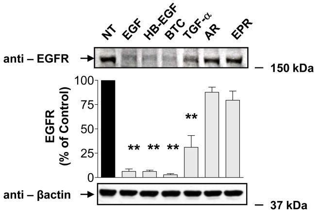
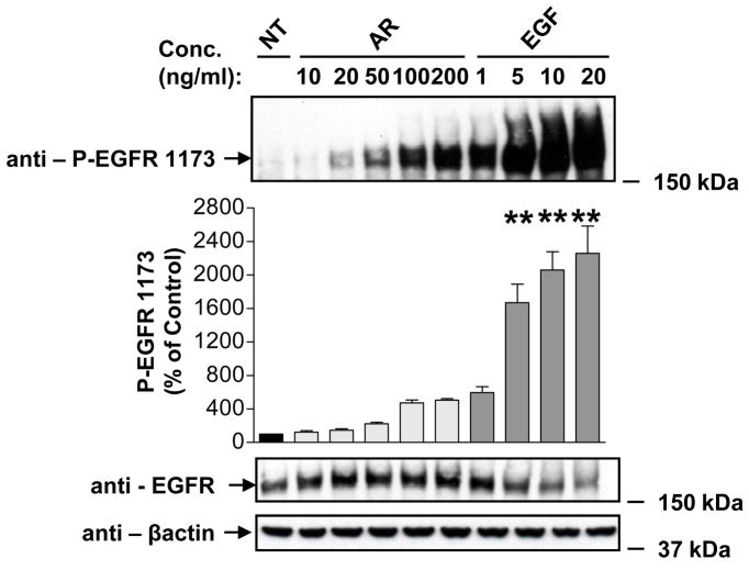
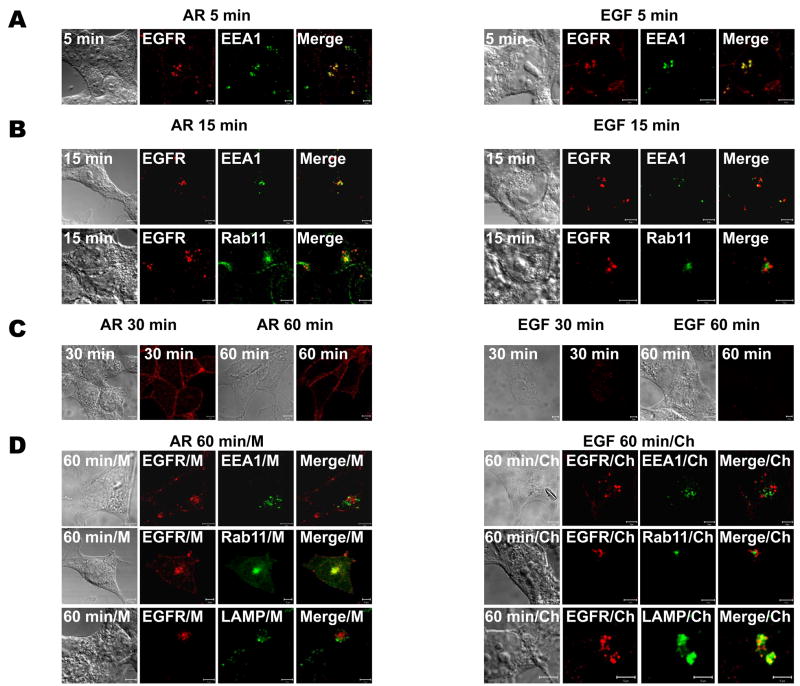
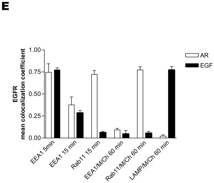
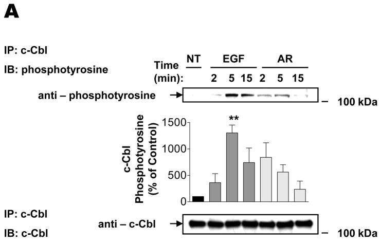
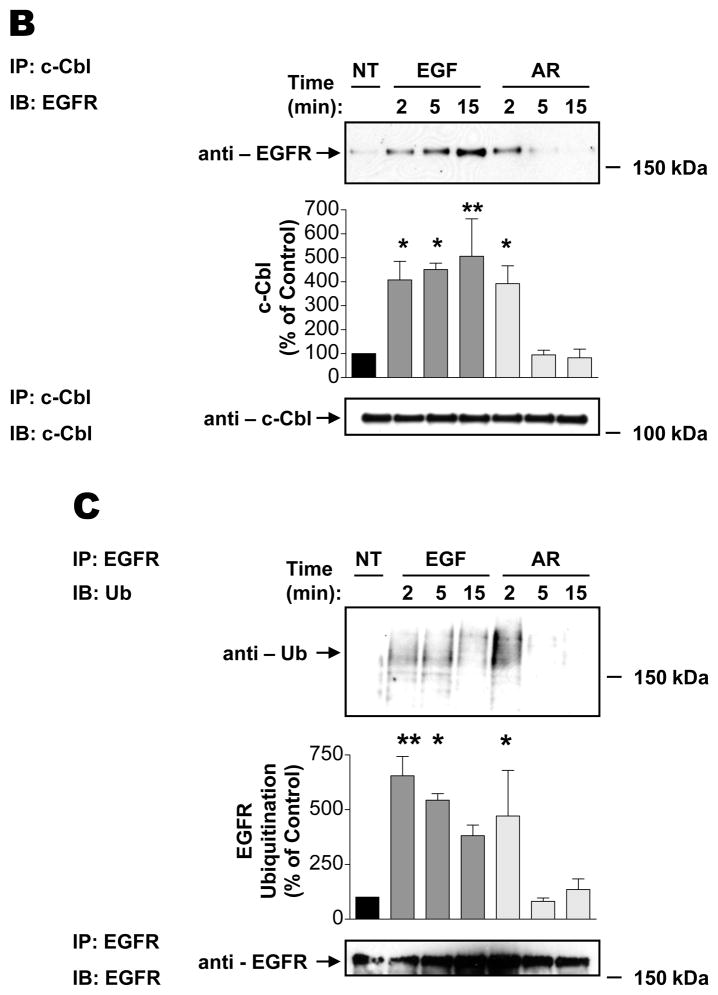
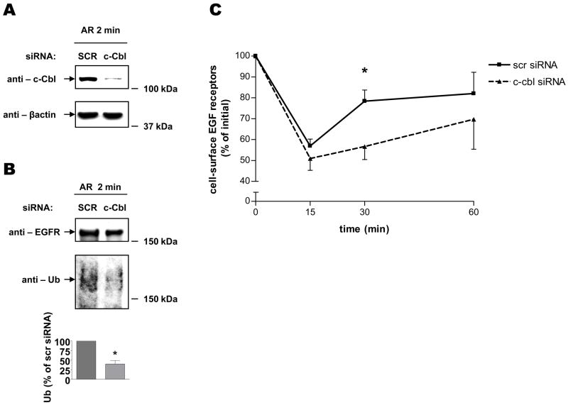
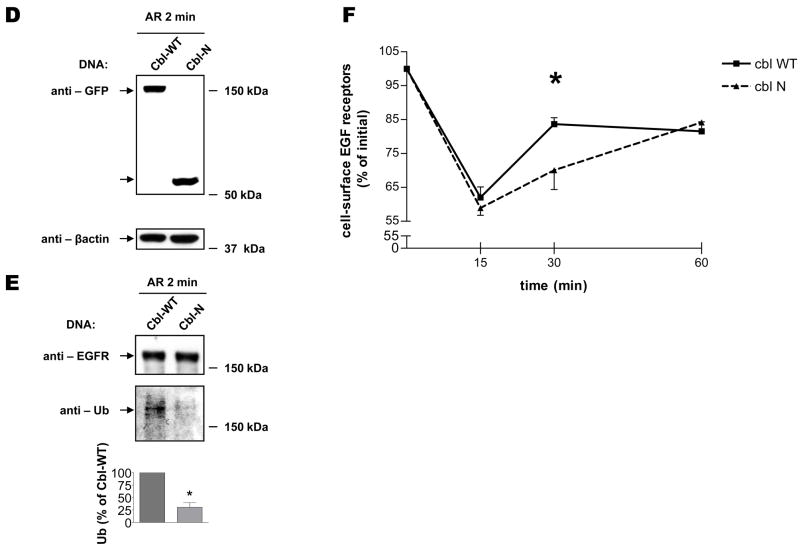
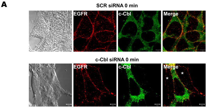
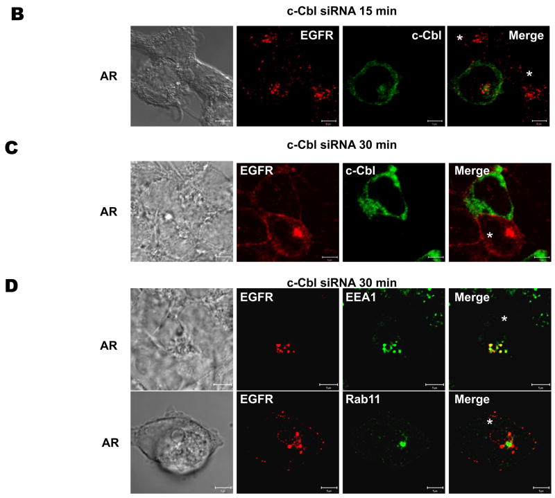
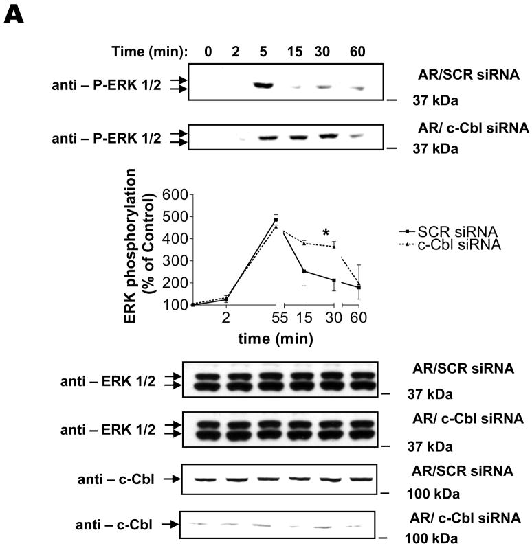
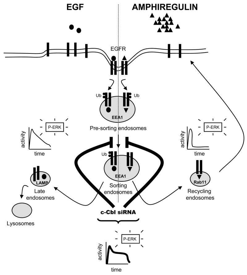
The intracellular processing of the epidermal growth factor receptor (EGFR) induced by epidermal growth factor (EGF) and transforming growth factor-alpha (TGF-alpha) has been studied meticulously, with the former resulting in EGFR degradation and the latter in EGFR recycling to the plasma membrane. However, little is known about how other EGF family growth factors affect the trafficking of the EGFR. Additionally, although both EGF and TGF-alpha have been shown to effectively induce initial c-Cbl (ubiquitin ligase)-mediated ubiquitination of the EGFR, limited information is available regarding the role of c-Cblin the trafficking and signaling of recycling EGFR. Thus, in this study, we investigated the roles of c-Cblin endogenous EGFR trafficking and signaling after stimulation with amphiregulin (AR). We demonstrated that a physiological concentration of AR induced recycling of the endogenous EGFR to the plasma membrane, which correlated closely with transient association of the EGFR with c-Cbl and transient EGFR ubiquitination. Most importantly, we used c-Cbl small interfering RNA (siRNA) duplexes and ac-Cbl dominant negative mutant to show that c-Cbl is critical for the efficient transition of the EGFR from early endosomes to a recycling pathway and that c-Cbl regulates the duration of extracellular signal regulated kinase 1/2 mitogen-activated protein kinase (ERK1/2 MAPK) phosphorylation. These data support novel functions of c-Cbl in mediating recycling of EGF receptors to the plasma membrane, as well as in mediating the duration of activation (transient vs sustained) of ERK1/2 MAPK phosphorylation.


Figures

FIGURE 1
FIGURE 1. Differential fates of EGFR induced by EGFR ligands
Serum-deprived HEK293 cells were treated at 37°C with vehicle (NT), 100 ng/ml EGF, HB-EGF, BTC, TGF-α, AR or EPR for 180 min, extracted with RIPA buffer and subsequently immunoblotted with anti-EGFR and anti-β-actin antibodies. Data shown are representative of three independent experiments. Results are mean ± S.E. (n=3), **, p <0.01 versus vehicle (NT).
FIGURE 2
FIGURE 2. Effects of AR and EGF on EGFR autophosphorylation
Serum-deprived HEK293 cells were treated at 37°C with vehicle (NT), 10, 20, 50, 100 and 200 ng/ml AR, or 1, 5, 10 and 20 ng/ml EGF for 2 min. After washing with ice-cold PBS, cells were extracted with RIPA buffer and cell lysates were immunoblotted with anti-phospho-EGFR Tyr-1173 antibody. Blots were then stripped and reprobed for total EGFR and β-actin to normalize for loading. Data shown are representative of three independent experiments. Results are mean ± S.E. (n=3), **, p <0.01 versus vehicle (NT).
FIGURE 3
FIGURE 3. Differential trafficking of EGFR induced by EGF and AR
Serum-deprived HEK293 cells were pre-treated without (panels A–C) or with 100 μM chloroquine (Ch) or 100 μM monensin (M) for 15 min (panel D), incubated on ice with 1 ng/ml EGF or 100 ng/ml AR for 45 min, washed free of unbound ligand, warmed, and exposed to pre-warmed ligand-free medium at 37°C for 5 (A), 15 (B), or 30 and 60 (CD) minutes. The cells then were fixed, stained with anti-EGFR antibody (visualized with Alexa Fluor 568-conjugated secondary antibody; red) and anti-EEA1, -Rab11 or -LAMP antibodies (visualized with Alexa Fluor 488-conjugated secondary antibody; green), and analyzed by confocal microscopy. Data shown are representative of three independent experiments. Yellow indicates co-localization. Bar, 5 μm. (E) The colocalizations between EGFR and EEA1, Rab11 or LAMP observed in panels A, B, and D were quantified using Zeiss LSM 510 META colocalization analysis software. The mean colocalization coefficients, averaged from at least three independent single cell images, represent pixel overlap between EGFR and EEA1, Rab11 or LAMP. The coefficients vary from 0 to 1, with 0 corresponding to non-overlapping images and 1 corresponding to 100% co-localization.
FIGURE 4
FIGURE 4. Effects of AR and EGF on EGFR ubiquitination and association with c-Cbl
Serum-deprived HEK293 cells were treated at 37°C with vehicle (NT), 1 ng/ml EGF or 100 ng/ml AR for 2, 5, 15 min. After washing with ice-cold PBS, cell lysates were (AB) subjected to immunoprecipitation with an antibody to c-Cbl, followed by immunoblotting with an antibody to phosphotyrosine or EGFR, or (C) subjected to immunoprecipitation with an antibody to EGFR, followed by immunoblotting with an antibody to ubiquitin. Blots were then stripped and reprobed for total c-Cbl or EGFR. Insets shown are representative of three independent experiments. Results are mean ± S.E. (n=3), *, p <0.05, **, p <0.01 versus vehicle (NT).
FIGURE 5
FIGURE 5. Role of c-Cbl in AR-induced EGFR recycling
Serum-deprived HEK293 cells, which had been transiently transfected with scrambled (SCR) or c-Cbl siRNA for 72 h (AC), or which had been transiently transfected with GFP-c-Cbl-WT or GFP-c-Cbl-N for 24 h (DF), were (A and D) treated with 100 ng/ml AR for 2 min, extracted with RIPA buffer, following which cell lysates were immunoblotted with an anti-c-Cbl, -GFP or –β-actin antibodies; (B and E) pre-incubated with sulfo-NHS-biotin for 30 min to label cell surface proteins and subsequently treated with 100 ng/ml AR for 2 min. After washing with ice-cold PBS, biotinylated proteins were analyzed by SDS-PAGE followed by immunoblotting with antibodies to EGFR and Ub; (C and F) incubated on ice with 100 ng/ml AR for 45 min, and subsequently incubated in pre-warmed ligand-free medium at 37°C for 0, 15, 30 and 60 min. The cells were then rinsed with ice-cold binding buffer, followed by a 7 minute long incubation with a low pH stripping buffer. The specific binding was determined by incubating cells for 90 min on ice with [125I]-EGF. The results are expressed as percentage of the original binding sites measured at 0 min. Data shown in panels A, B D, and E are representative of three independent experiments. Results in panel C and F are mean ± S.E. of three separate experiments, *, p < 0.05 versus scrambled siRNA.
FIGURE 6
FIGURE 6. Role of c-Cbl in AR-induced EGFR trafficking
Serum-deprived HEK293 cells, which had been transiently transfected with scrambled (SCR) or c-Cbl siRNA for 72 h, were incubated on ice with 100 ng/ml AR for 45 min, washed free of unbound ligand, and subsequently incubated in pre-warmed ligand-free medium at 37°C for 0 (A), 15 (B), and 30 (C) min. The cells then were fixed, stained with anti-EGFR (visualized with Alexa Fluor 568-conjugated secondary antibody; red) and anti-c-Cbl (visualized with Alexa Fluor 488-conjugated secondary antibody; green) antibodies, and analyzed by confocal microscopy. Fields were chosen to show simultaneously cells in which c-Cbl was depleted and cells in which c-Cbl was not, in order to facilitate direct comparisons. White asterisks show positions of c-Cbl-depleted cells. (D) These micrographs show the intracellular localization of EGFR (visualized with Alexa Fluor 568-conjugated secondary antibody; red), in serum-deprived HEK293 cells, which had been transiently transfected with scrambled (SCR) or c-Cbl siRNA for 72 h, 30 minutes after a synchronized pulse with AR. Dual labeling was performed with markers of subcellular organelles, i.e. early endosomes and the perinuclear recycling compartment (visualized with anti-EEA1 and -Rab11 antibodies, respectively, and Alexa Fluor 488-conjugated secondary antibody; green). White asterisks show positions of c-Cbl-depleted cells. Yellow indicates co-localization. Bar, 5 μm. Data shown are representative of at least three independent experiments.
FIGURE 7
FIGURE 7. Role of c-Cbl in AR-induced MAPK activation
Serum-deprived HEK293 cells, which had been transiently transfected with scrambled (SCR) or c-Cbl siRNA for 72 h, were incubated at 37°C with 100 ng/ml AR for 0, 2, 5, 15, 30, and 60 min. After washing with ice-cold PBS, cells were extracted with RIPA buffer, following which cell lysates were immunoblotted with anti-phospho ERK1/2. Blots then were stripped and reprobed for total ERK1/2 or c-Cbl. Results are mean ± S.E. (n=3), *, p < 0.05 versus scrambled siRNA. Insets shown are representative of three independent experiments.
FIGURE 8
FIGURE 8. Roles of c-Cbl in EGFR recycling and signaling
Binding of AR (triangles; right-side of scheme) or EGF (ovals; left-side of scheme) to the EGFR triggers receptor endocytosis. Following internalization into early pre-sorting endosomes (EEA1-positive), the AR- or EGF-stimulated EGFR is sorted into recycling endosomes (Rab11-positive), or to late endosomes and lysosomes (LAMP-positive), respectively. Differential trafficking of EGFR correlates with differential patterns of ERK activation, i.e. unlike AR, which causes transient phosphorylation of ERK, EGF results in much more persistent activation of ERK. AR induces transient ubiquitination of EGFR (small circles, Ub), whereas EGF induces more sustained ubiquitination. Regardless of EGFR ligand, c-Cbl regulates exit into both recycling and degradative EGFR trafficking pathways. In the absence of c-Cbl, the disrupted sorting of EGFR causes receptor retention within the early endosomes, which consequently is associated with more sustained phosphorylation of ERK.

References

    1. Wiley HS. Trafficking of the ErbB receptors and its influence on signaling. Exp Cell Res. 2003;284:78–88. - PubMed
    1. Ebner R, Derynck R. Epidermal growth factor and transforming growth factor-alpha: differential intracellular routing and processing of ligand-receptor complexes. Cell Regul. 1991;2:599–612. - PMC - PubMed
    1. Longva KE, Blystad FD, Stang E, Larsen AM, Johannessen LE, Madshus IH. Ubiquitination and proteasomal activity is required for transport of the EGF receptor to inner membranes of multivesicular bodies. J Cell Biol. 2002;156:843–854. - PMC - PubMed
    1. Stern KA, Place TL, Lill NL. EGF and amphiregulin differentially regulate Cbl recruitment to endosomes and EGF receptor fate. Biochem J. 2008;410:585–594. - PMC - PubMed
    1. Di Fiore PP, Gill GN. Endocytosis and mitogenic signaling. Curr Opin Cell Biol. 1999;11:483–488. - PubMed

Publication types