Review
. 2009 Feb 20;4(2):93-107.
doi: 10.1021/cb8002946.

Ribosomal translocation: one step closer to the molecular mechanism

Affiliations
Review

Ribosomal translocation: one step closer to the molecular mechanism

Shinichiro Shoji et al. ACS Chem Biol. .

Abstract

Protein synthesis occurs in ribosomes, the targets of numerous antibiotics. How these large and complex machines read and move along mRNA have proven to be challenging questions. In this Review, we focus on translocation, the last step of the elongation cycle in which movement of tRNA and mRNA is catalyzed by elongation factor G. Translocation entails large-scale movements of the tRNAs and conformational changes in the ribosome that require numerous tertiary contacts to be disrupted and reformed. We highlight recent progress toward elucidating the molecular basis of translocation and how various antibiotics influence tRNA-mRNA movement.


Figures

Figure 1
Figure 1
Scheme for a translation elongation cycle. Each cycle of translation elongation is composed of three major steps: decoding, peptidyl transfer, and translocation. In decoding, aminoacyl-tRNA (aa-tRNA) is delivered to the A site as part of a ternary complex with EF-Tu and GTP. This is followed by rapid and functionally irreversible transfer of the peptidyl group from P-tRNA to A-tRNA. EF-G·GTP then catalyzes translocation, the coupled movement of tRNA and mRNA in the ribosome. Deacylated tRNA dissociates from the E site before or during the next round of elongation.
Figure 2
Figure 2
tRNA–ribosome interactions in A, P, and E sites. a) A model of a ribosomal complex containing mRNA and tRNAs in all three sites. b) Interactions at the 3′ CCA ends of A-tRNA and P-tRNA. c) Interactions at the 30S A site. d) Interactions of P-tRNA with H69 of 23S rRNA and ribosomal proteins L5 and S13. e) Type I/II base triples at A1339 and G1338 of 16S rRNA in the 30S P site and the “gate” formed by the A790 loop and the 1338–1339 loop. f) Interactions between A76 of tRNA and 23S rRNA. g) Interactions between the L1 stalk and E-tRNA. h) Interactions between E-tRNA and ribosomal protein S7. Images in panels a and c–h were generated from crystal structures of Thermus thermophilus ribosomes containing A-, P-, E-tRNA, and mRNA (PDB IDs: 2HGP, 2HGQ, 2J00, and 2J01) (14, 125). An image in panel b was generated from a structure of the Halo-arcula marismortui 50S subunit with A- and P-site substrates (PDB ID: 1KQS) (126). 23S rRNA and 16S rRNA are shown in gray and cyan, respectively. E. coli numbering of rRNA nucleotides is used throughout.
Figure 3
Figure 3
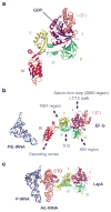
Structure of EF-G. a) A crystal structure of T. thermophilus EF-G·GDP (PDB ID: 2EFG) (52). Domain III is partially disordered and thus not fully visible. b) A structural model of EF-G based on cryo-EM reconstructions of complexes containing EF-G·GDPNP and P/E-tRNA (PDB ID: 2OM7) (64). Notable interactions of EF-G with ribosomes are shown by arrows. Ribosomal components in the 50S and 30S subunits are colored in gray and blue, respectively. c) A structural model of LepA based on cryo-EM reconstructions of complexes containing LepA·GDPNP, P-tRNA, and A/L-tRNA (PDB ID: 3DEG) (116).
Figure 4
Figure 4
Structural rearrangements in the 30S subunit. Connected dots represent movement of phosphorus atoms in 16S rRNA during the RSR (panel a) or the head swiveling (panel b). Images were generated from available cryo-EM (PDB ID: 1P87, 1P86, 1P6G, and 1P85) (127) and crystal structures (PDB ID: 2AVY, 2AW7, 2AW4, and 2AWB) (24).
Figure 5
Figure 5
Kinetic model for EF-G-dependent translocation.
Figure 6
Figure 6
Chemical structures of antibiotics that affect tRNA–mRNA movement.
Figure 7
Figure 7
Structural rearrangements in h44 of 16S rRNA induced by aminoglycosides. Conformations of 16S rRNA nucleotides 1491–1494 in E. coli 70S ribosomes in the absence (panel a) or presence of neomycin (panel b) or hygromycin B (panel c) are shown. Paromomycin and gentamicin induce the same h44 conformation as neomycin (91). Structures were adopted from crystal structures of E. coli 70S ribosomes in complex with various antibiotics (PDB ID: 2AVY, 2QAL, and 3DF1) (24, 91).
Figure 8
Figure 8
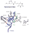
Sparsomycin interacts with the 3′ end of peptidyl-tRNA in the 50S P site. Interactions at the peptidyl transferase center of the H. marismortui 50S subunit containing P-site substrate and sparsomycin (PDB ID: 1VQ8) (117). 23S rRNA is colored in gray. E. coli numbering is used throughout.
Figure 9
Figure 9
A possible mechanism for sparsomycin-dependent translocation. Sparsomycin (Sps) may promote efficient translocation by stabilizing the unlocked POST state (POST*) relative to the unlocked hybrid PRE state (PRE(HS)*). This model postulates that during translocation unlocking structural rearrangements would allow Brownian movement of tRNA in a complex that rapidly fluctuates between PRE(HS)* and POST*. Complexes in POST* would then be locked in the POST state by reverse structural rearrangements (relocking). Size of the letters underneath each complex infers relative stability of the complex.

References

    1. Gromadski KB, Rodnina MV. Kinetic determinants of high-fidelity tRNA discrimination on the ribosome. Mol Cell. 2004;13:191–200. - PubMed
    1. Ogle JM, Ramakrishnan V. Structural insights into translational fidelity. Annu Rev Biochem. 2005;74:129–177. - PubMed
    1. Palade GE. A small particulate component of the cytoplasm. J Biophys Biochem Cytol. 1955;1:59. - PMC - PubMed
    1. Ban N, Nissen P, Hansen J, Moore PB, Steitz TA. The complete atomic structure of the large ribosomal subunit at 2.4 Å resolution. Science. 2000;289:905–920. - PubMed
    1. Carter AP, Clemons WM, Brodersen DE, Morgan-Warren RJ, Wimberly BT, Ramakrishnan V. Functional insights from the structure of the 30S ribosomal subunit and its interactions with antibiotics. Nature. 2000;407:340–348. - PubMed

MeSH terms