. 2009 Jan 27:9:16.
doi: 10.1186/1471-2180-9-16.

Ultrastructure and molecular phylogeny of Calkinsia aureus: cellular identity of a novel clade of deep-sea euglenozoans with epibiotic bacteria

Affiliations

Ultrastructure and molecular phylogeny of Calkinsia aureus: cellular identity of a novel clade of deep-sea euglenozoans with epibiotic bacteria

Naoji Yubuki et al. BMC Microbiol. .

Abstract

Background: The Euglenozoa is a large group of eukaryotic flagellates with diverse modes of nutrition. The group consists of three main subclades - euglenids, kinetoplastids and diplonemids--that have been confirmed with both molecular phylogenetic analyses and a combination of shared ultrastructural characteristics. Several poorly understood lineages of putative euglenozoans live in anoxic environments, such as Calkinsia aureus, and have yet to be characterized at the molecular and ultrastructural levels. Improved understanding of these lineages is expected to shed considerable light onto the ultrastructure of prokaryote-eukaryote symbioses and the associated cellular innovations found within the Euglenozoa and beyond.

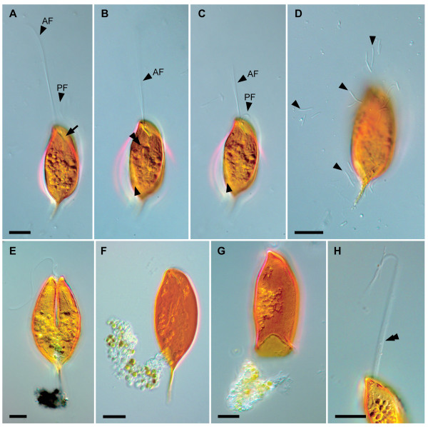
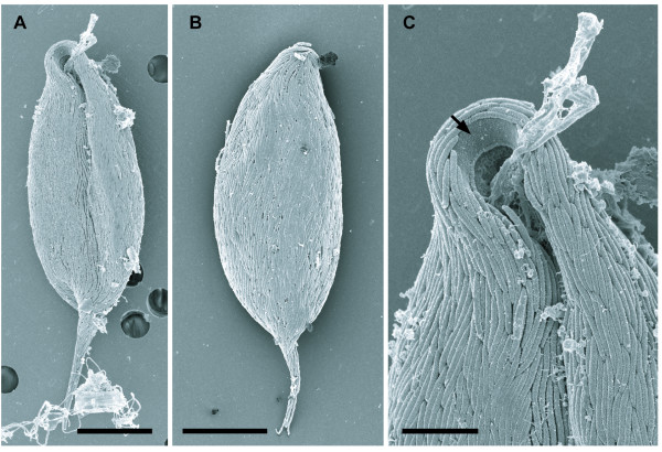
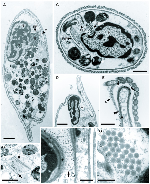
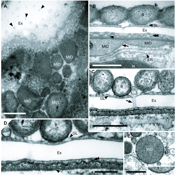
Results: We collected Calkinsia aureus from core samples taken from the low-oxygen seafloor of the Santa Barbara Basin (580 - 592 m depth), California. These biflagellates were distinctively orange in color and covered with a dense array of elongated epibiotic bacteria. Serial TEM sections through individually prepared cells demonstrated that C. aureus shares derived ultrastructural features with other members of the Euglenozoa (e.g. the same paraxonemal rods, microtubular root system and extrusomes). However, C. aureus also possessed several novel ultrastructural systems, such as modified mitochondria (i.e. hydrogenosome-like), an "extrusomal pocket", a highly organized extracellular matrix beneath epibiotic bacteria and a complex flagellar transition zone. Molecular phylogenies inferred from SSU rDNA sequences demonstrated that C. aureus grouped strongly within the Euglenozoa and with several environmental sequences taken from low-oxygen sediments in various locations around the world.

Conclusion: Calkinsia aureus possesses all of the synapomorphies for the Euglenozoa, but lacks traits that are specific to any of the three previously recognized euglenozoan subgroups. Molecular phylogenetic analyses of C. aureus demonstrate that this lineage is a member of a novel euglenozoan subclade consisting of uncharacterized cells living in low-oxygen environments. Our ultrastructural description of C. aureus establishes the cellular identity of a fourth group of euglenozoans, referred to as the "Symbiontida".


Figures

Figure 1
Figure 1
Differential interference contrast images of the living cell of Calkinsia aureus. The micrographs show the distinctively orange color of the cell, two flagella, epibiotic bacteria and ingested material. A. Anterior flagellum (AF) and the posterior flagellum (PF) inserted into an anterior opening (arrow). The ingested material is present in the middle and posterior regions of the cell. B. Surface striations (arrowhead) and a longitudinal rod-like structure (double arrowhead) indicative of a feeding apparatus. C. AF and PF emerging from the anterior opening. The arrowhead shows striation on the surface of the cell. D. Bacteria (arrowheads) that have disassociated with C. aureus. E. A cell undergoing division showing a longitudinal cleavage furrow starts from the anterior end. The ingested material is present in the middle and posterior regions of the cell. F. Clear cytoplasm extruded from posterior of the cell. G. Bright orange extracellular matrix. H. Bundle of extrusomes (double arrowhead) that have been discharged from extrusomal pocket through the anterior opening. (bars = 10 μm, A-C at same scale).
Figure 2
Figure 2
Scanning electron micrographs (SEM) of Calkinsia aureus. A. The ventral side of C. aureus showing the anterior opening, a longitudinal groove and epibiotic bacteria. B. The dorsal side of the C. aureus showing the epibiotic bacteria. (A, B bars = 10 μm). C. High magnification SEM of the anterior vestibular opening showing the absence of epibiotic bacteria on the extracellular matrix (arrow). (bar = 3 μm).
Figure 3
Figure 3
Transmission electron micrographs (TEM) showing the general morphology of Calkinsia aureus. A. Sagittal TEM showing the nucleus (N) with condensed chromatin and a conspicuous nucleolus (Nu), a battery of extrusomes (E), the vestibulum (V) located on the dorsal side of the cell, ingested material and epibiotic bacteria on the extracellular matrix. The extrusomal pocket (EP) branched from the vestibulum (V) (bar = 4 μm). B. Ingested material containing diatom frustules (arrow). (bar = 2 μm). C. Cross section of the cell through the nucleus (N), the battery of extrusomes (E), the flagellar pocket (FLP) and the feeding pocket (FdP). (bar = 2 μm). D. High magnification view through the vestibulum (V) that is opened on the ventral side of the cell. E. High magnification view through the anterior opening showing the termination of the extracellular matrix (double arrowhead) and fine somatonemes (S) or hair-like structures on the perforated matrix (arrows) that is not covered with epibiotic bacteria. The arrowhead indicates the supportive microtubular sheet that lines the inside of the cytostome and turns along the cell surface. (D, E, bars = 1 μm). F. Hairs (arrow) on the wall of the vestibulum (V). (bar = 1 μm). G. Cross section showing the battery of tubular extrusomes (E). (bar = 2 μm).
Figure 4
Figure 4
Transmission electron micrographs (TEM) showing the surface ultrastructure of Calkinsia aureus. A. Tangential TEM section showing conduit-like perforations (arrowheads) embedded within the extracellular matrix (Ex), an array of microtubules, and mitochondrion-derived organelles (MtD). (bar = 1 μm). B. Mitochondrion-derived organelles (MtD) with two membranes (arrow) above the ER. The convoluted appearance of the cell plasma membrane (double arrowhead) and a longitudinal view of a microtubule (arrowhead) are also shown. A glycocalyx (GL) covers the surface of the extracellular matrix (Ex). C. Transverse TEM showing the epibiotic bacteria (B), the glycocalyx (GL), a conduit-like perforation (arrow) through the extracellular matrix (Ex) and the underlying sheet of microtubules (B, C, bars = 500 nm). D. High magnification view showing the epibiotic bacteria (B), the glycocalyx (GL), the extracellular matrix (Ex), the cell plasma membrane (double arrowhead), and the double-layered structure (arrowhead; derived from the dorsal lamina) beneath a sheet of inter-connected microtubules (bar = 200 nm). E. Mitochondrion-derived organelles (MtD) (bar = 500 nm). Inset: High magnification TEM showing the two membranes that surround the mitochondrion-derived organelles (width of inset = 400 nm).
Figure 5
Figure 5
Diagram of the cell surface of Calkinsia aureus. The diagram shows epibiotic bacteria (B), the glycocalyx (GL), the perforated extracellular matrix (Ex), the host cell plasma membrane (double arrowhead), the linked microtubules (LMt), the double-layered structure (arrowhead), mitochondrion-derived organelles (MtD) and cisternae of endoplasmic reticulum (ER).
Figure 6
Figure 6
Transmission electron micrographs (TEM) showing paraxonemal rods in the flagella, the flagellar transition zone and the basal bodies of Calkinsia aureus. A. Longitudinal section of the dorsal flagellum (DF) showing the flagellar transition zone and the dorsal basal body (DB) (bar = 500 nm). B-J. Non-consecutive serial sections through the DF (B), the flagellar transition zone (C-G), and the DB (H-J) as viewed from anterior end (images at same scale, bar = 200 nm). B. Section showing the 9+2 configuration of axonemal microtubules and the tubular paraxonemal rod (arrow) in the DF. C. Section showing termination of central microtubules and the 9+0 configuration of axonemal microtubules. D. Section showing the transition zone through an outer concentric ring associated with nine electron dense globules inside of each doublet and faint spokes that extend inward from the each globule (see L for a diagram of this micrograph). E. Section through the nine radial connectives (arrowhead) that extend outward from each doublet to the flagellar membrane. F. Section showing the radial connectives that extend outward toward the flagellar membrane, the spokes that extend inward from the microtubular doublets, the central electron dense hub, and inner concentric rings (see M for the diagram of this micrograph). G. Section showing the electron dense hub and inner and outer concentric rings, and the absence of radial connectives. H. A section at the level of the insertion of the DF. The transitional fibers (double arrowheads) extending from the microtubular triplets of the DB are shown. I. Section through the area just below the distal boundary of the DB. The transitional fibers (double arrowheads) connect to each microtubular triplet. J. Section through the proximal region of the DB showing the cartwheel structure. K. View through the paraxonemal rod of the ventral flagellum (VF) (bar = 500 nm). L. Diagram of the level of D showing faint spokes (a) that extend inward from each globule, an outer concentric ring (b) and nine electron dense globules (c). M. Diagram of the level of F showing spokes (a), an outer concentric ring (b), nine electron dense globules (c), an electron dense hub (d), an inner concentric ring (e) and radial connectives (f).
Figure 7
Figure 7
Transmission electron micrographs (TEM) showing the organization of microtubular roots that originate from the dorsal and ventral basal bodies (DB and VB, respectively). Those are viewed from the anterior end (A-F at same scale, bar = 500 nm). A. The proximal region of the basal bodies close to the cartwheel structure. The dorsal root (DR) originates from the DB; the intermediate root (IR) and the ventral root (VR) extend from the VB. A dorsal lamina (DL) attaches to the dorsal side of the DR; the right fiber (RF) is close to the ventral side of the VR. B. Section showing the right fiber (RF), the IR-associated lamina (IL), a left fiber (LF) and an intermediate fiber (IF) associated with the VB. The arrow points to the connective fiber between the DB and the VB. Dense fibrils (double arrowhead) extend to the right side of each microtubule of the intermediate root (IR). C. Section through the middle part of the DB and the VB. D. Section through the insertion of the flagella. E. Section through the flagellar transition zone showing the extension of the DL and disorganization of the VF. F. Section showing the linked microtubules (LMt) associated with the dorsal lamina (DL) and the ventral root (VR). G. High magnification view of proximal area of the two basal bodies, the DB and the VB, of A showing the cartwheel structure and the dorsal lamina (DL) on the dorsal side of the dorsal root (DR). The double arrowhead indicates the fibril from each microtubule of the IR. H. High magnification view of right wall of the pocket of F showing the LMt and the DL. I. High magnification view of the IR of D showing the relationship among the IR, IL and IF. J. High magnification view of the IR at the level of E showing the IR and IL. K. High magnification view of the IR and IL. L. High magnification view of the VR in E. (G-L, bars = 200 nm).
Figure 8
Figure 8
Transmission electron micrographs (TEM) of Calkinsia aureus showing the feeding apparatus. The ventral flagellum was disorganized in all sections (A-D at same scale, bar = 1 μm; E-G at same scale, bar = 1 μm). A. Section showing the oblique striated fibrous structure (OSF) and the VR along the wall of the flagellar pocket (FLP). Arrow points out the LMt and the DL. B. Section through the congregated globular structure (CGS), the OSF and the feeding pocket (FdP). The VR extends to the right. The arrow points out the LMt and the DL, which extend from the VR to the IR and support the dorsal half of the FLP. C. Section showing the VR over the CGS. Arrows show the LMt and DL. D. The VR crosses over the CGS and extends to right side of the FdP. Most of the wall of the FLP is supported by the LMt and DL (arrows). E. A striated fiber (double arrowhead) supports the left side of the FdP and extends from the left side of the CGS. Arrows indicate the extension of the LMt and DL. F. Section through the beginning of the vestibulum (V) and the striated fiber (double arrowhead). G. The V is enlarged and the CGS remains at both sides of the FdP. H. High magnification of FdP. I. Tangential TEM section showing the VR with an electron dense fiber along the feeding pocket and a tomentum (T) of fine hairs. J. Longitudinal section through the CGS and the OSF. Six ventral root microtubules embedded within the electron dense fibers (arrowheads). K. High magnification view of the VR supporting the FdP shown in F. Double arrowhead indicates the striated fiber and the six arrowheads indicate the electron dense fibers of the VR. (H-K, bars = 500 nm).
Figure 9
Figure 9
Diagram of the cell (A), the flagellar apparatus (B) and the feeding apparatus (C) of Calkinsia aureus based on serial TEM sections. A. Illustration of the cell viewed from the left side; arrow marks the extrusomal pocket. Boxes B and C indicate the plane of view shown in Figures B and C, respectively. B. Illustration of the flagellar apparatus as viewed from left side. C. Illustration of the feeding apparatus as viewed from anterior-ventral side. The double arrowhead marks the striated fiber along the feeding pocket (FdP). Note DL, IF, IL, LF, LMt, and RF are not shown on this diagram for clarity.
Figure 10
Figure 10
Phylogenetic position of Calkinsia aureus within eukaryotes as inferred from SSU rRNA gene sequences. Maximum likelihood (ML) analysis of 38 taxa sampled from phylogenetically diverse eukaryotes. This tree is rooted with opisthokont sequences. ML bootstrap values greater than 50% are shown. Thick branches indicate Bayesian posterior probabilities over 0.95. GenBank accession numbers of the sequences analyzed are shown in parentheses.
Figure 11
Figure 11
Phylogenetic position of Calkinsia aureus within euglenozoans as inferred from SSU rRNA gene sequences. Maximum likelihood (ML) analysis of 35 taxa focusing on the position of Calkinsia aureus within the Euglenozoa clade. Two jakobids, Andalucia incarcerata and A. godoyi, are used as outgroups in this analysis. ML bootstrap values greater than 50% are shown. Thick branches indicate Bayesian posterior probabilities over 0.95. Ba, bacteriotroph; Eu, eukaryotroph; Ph, phototroph. GenBank accession numbers of the sequences analyzed are shown in parentheses.

Similar articles

Cited by

References

    1. Keeling PJ, Burger G, Durnford DG, Lang BF, Lee RW, Pearlman RE, Roger AJ, Gray MW. The tree of eukaryotes. Trends Ecol Evol. 2005;20:670–676. - PubMed
    1. Yoon HS, Grant J, Tekle YI, Wu M, Chaon BC, Cole JC, Logsdon JM Jr, Patterson DJ, Bhattacharya D, Katz LA. Broadly sampled multigene trees of eukaryotes. BMC Evol Biol. 2008;8:14. - PMC - PubMed
    1. Adl SM, Simpson AGB, Farmer MA, Andersen RA, Anderson OR, Barta JR, Bowser SS, Brugerolle G, Fensome RA, Fredericq S, James TY, Karpov S, Kugrens P, Krug J, Lane CE, Lewis LA, Lodge J, Lynn DH, Mann DG, McCourt RM, Mendoza L, Moestrup Ø, Mozley-Standridge SE, Nerad TA, Shearer CA, Smirnov AV, Spiegel FW, Taylor MF. The new higher level classification of eukaryotes with emphasis on the taxonomy of protists. J Eukaryot Microbiol. 2005;52:399–451. - PubMed
    1. Adl SM, Leander BS, Simpson AGB, Archibald JM, Anderson OR, Bass D, Bowser SS, Brugerolle G, Farmer MA, Karpov S, Kolisko M, Lane CE, Lodge DJ, Mann DG, Meisterfeld R, Mendoza L, Moestrup Ø, Mozley-Standridge SE, Smirnov AV, Spiegel F. Diversity, nomenclature, and taxonomy of protists. Syst Biol. 2007;56:684–689. - PubMed
    1. Cavalier-Smith T. Kingdom protozoa and its 18 phyla. Microbiol Rev. 1993;57:953–994. - PMC - PubMed

Publication types

LinkOut - more resources