. 2009 Jan 27;10(1):6.
doi: 10.1186/1465-9921-10-6.

Impact of interleukin-6 on hypoxia-induced pulmonary hypertension and lung inflammation in mice

Affiliations

Impact of interleukin-6 on hypoxia-induced pulmonary hypertension and lung inflammation in mice

Laurent Savale et al. Respir Res. .

Abstract

Background: Inflammation may contribute to the pathogenesis of various forms of pulmonary hypertension (PH). Recent studies in patients with idiopathic PH or PH associated with underlying diseases suggest a role for interleukin-6 (IL-6).

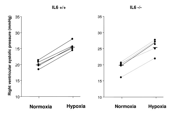
Methods: To determine whether endogenous IL-6 contributes to mediate hypoxic PH and lung inflammation, we studied IL-6-deficient (IL-6-/-) and wild-type (IL-6+/+) mice exposed to hypoxia for 2 weeks.

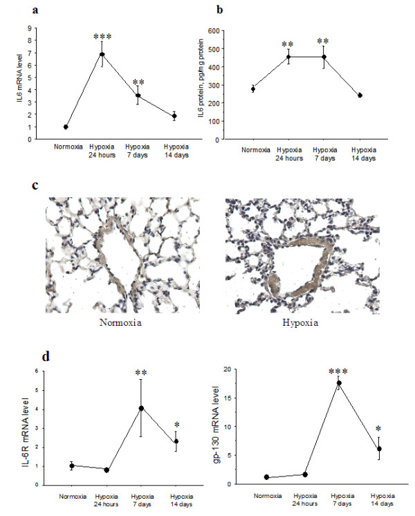
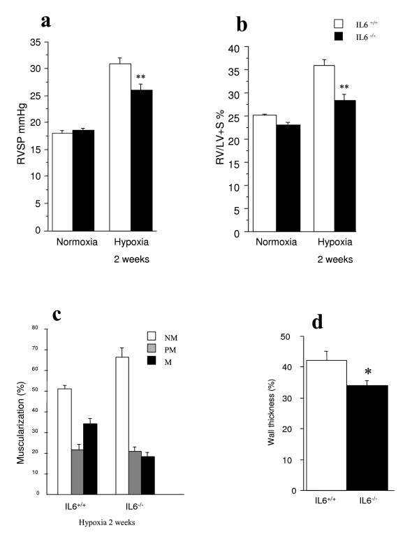
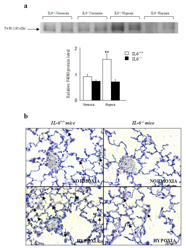
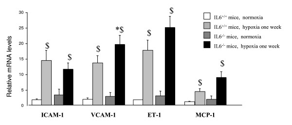
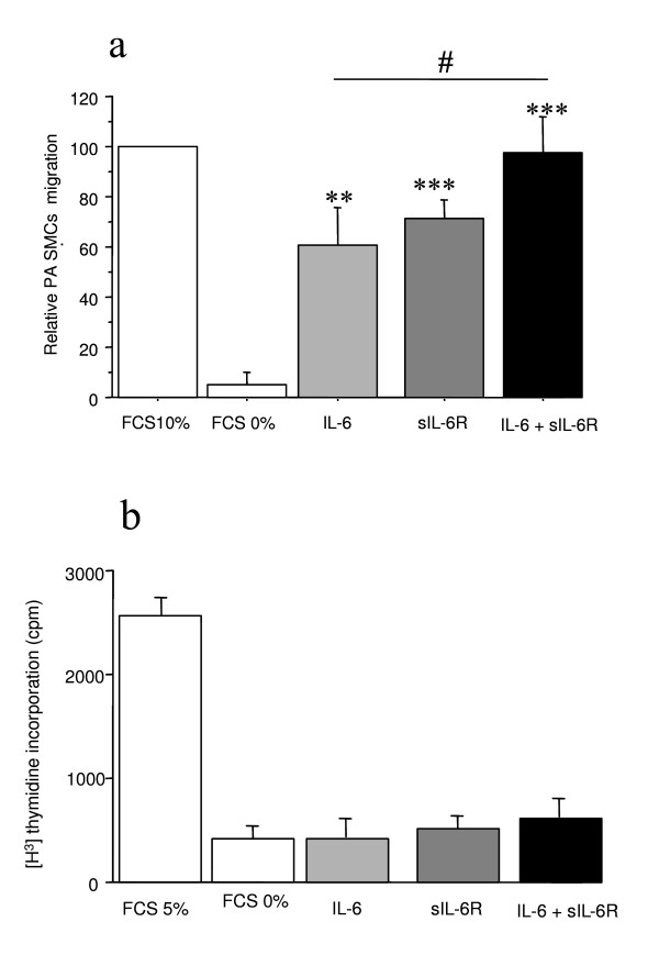
Results: Right ventricular systolic pressure, right ventricle hypertrophy, and the number and media thickness of muscular pulmonary vessels were decreased in IL-6-/- mice compared to wild-type controls after 2 weeks' hypoxia, although the pressure response to acute hypoxia was similar in IL-6+/+ and IL-6-/- mice. Hypoxia exposure of IL-6+/+ mice led to marked increases in IL-6 mRNA and protein levels within the first week, with positive IL-6 immunostaining in the pulmonary vessel walls. Lung IL-6 receptor and gp 130 (the IL-6 signal transducer) mRNA levels increased after 1 and 2 weeks' hypoxia. In vitro studies of cultured human pulmonary-artery smooth-muscle-cells (PA-SMCs) and microvascular endothelial cells revealed prominent synthesis of IL-6 by PA-SMCs, with further stimulation by hypoxia. IL-6 also markedly stimulated PA-SMC migration without affecting proliferation. Hypoxic IL-6-/- mice showed less inflammatory cell recruitment in the lungs, compared to hypoxic wild-type mice, as assessed by lung protein levels and immunostaining for the specific macrophage marker F4/80, with no difference in lung expression of adhesion molecules or cytokines.

Conclusion: These data suggest that IL-6 may be actively involved in hypoxia-induced lung inflammation and pulmonary vascular remodeling in mice.


Figures

Figure 1
Figure 1
Hemodynamic response to acute hypoxia in IL-6+/+ and IL-6-/- mice. Individual and mean (horizontal line) right ventricular systolic pressures (RSVP) in normoxic IL-6+/+ and IL-6-/- mice under ventilation with room air (normoxia) and after 5 min of ventilation with a hypoxic gas mixture (hypoxia). The increase in RVSP induced by acute exposure to 8%O2 did not differ between wild-type and IL-6-/- mice.
Figure 2
Figure 2
expression and immunolocalization of interleukin-6 in lungs from IL-6+/+ mice after hypoxia exposure. IL-6 mRNA levels in total lung tissue determined by real-time quantitative RT-PCR (a) and protein levels assessed by ELISA (b). Each point is the mean ± SEM of at least 8 determinations after exposure to 10% O2 for 24 hours, 1 week, or 2 weeks. **P < 0.01, ***P < 0.001 compared with values in normoxic mice. IL-6 immunostaining in lung sections from IL-6+/+ mice under normoxia (c, left panel) and after hypoxia exposure for 7 days (c, right panel). Strong IL-6 immunostaining is visible in vessel walls from the animal exposed to hypoxia (arrows). IL-6R and gp-130 RNA expression in total lung tissue from IL-6+/+ mice exposed to hypoxia (d). Each point is the mean ± SEM of at least 8 determinations after exposure to 10% O2 for 24 hours, 1 week, or 2 weeks. *P < 0.05, **P < 0.01, ***P < 0.001 compared to values in normoxic animals.
Figure 3
Figure 3
Development of hypoxic pulmonary hypertension and vascular remodeling in IL-6+/+ and IL-6-/- mice. Right ventricular systolic pressure (RVSP) (a) and weights of the right ventricle/left ventricle + septum (Fulton's index) (b) in IL-6+/+ and IL-6-/- mice exposed to normoxia or 10% O2 for 2 weeks. **P < 0.01 compared to IL-6+/+ mice under similar conditions. Percentage of muscularized vessels from wild-type IL-6+/+ and IL-6-/- mice (c). Twenty to thirty intraacinar vessels were examined in each lung from mice of each genotype after exposure to hypoxia for 2 weeks. Percentages of nonmuscular (NM), partially muscular (PM), and fully muscular (M) vessels differed significantly between IL-6+/+ and after 2 weeks of hypoxia (P < 0.05). Normalized wall thickness measured in fully muscular arteries in lungs from IL-6-/- and IL-6+/+ mice exposed to hypoxia for 2 weeks (d). *P < 0.05 compared to IL-6+/+ mice exposed to hypoxia for 2 weeks.
Figure 4
Figure 4
Lung macrophages recruitment under hypoxic condition in IL-6+/+ and IL-6-/- mice. Lung F4/80 protein levels assessed by Western blotting in IL-6+/+ mice and IL-6-/-mice after normoxia or hypoxia (n = 5 in each group) (a). Each bar is the mean ± SEM. *P < 0.05 compared to IL-6+/+ mice exposed to hypoxia of the same duration. Lung macrophage recruitment illustrated by representative photomicrographs showing F4/80 immunostaining in lung sections from IL-6+/+ and IL-6-/- mice under normoxia and hypoxia (b). Macrophages are shown by arrows.
Figure 5
Figure 5
Lung expression of ICAM-1, VCAM-1, ET-1 and MCP-1 mRNAs in IL-6-/- and IL-6+/+ during normoxic and hypoxic conditions. Levels of ICAM-1, VCAM-1, ET-1, and MCP-1 mRNAs in lung tissue from IL-6+/+ and IL-6-/- mice after normoxia or 1 week of hypoxia. Each bar is the mean ± SEM (n = 5 in each group). *P < 0.05 and **P < 0.01 compared with corresponding values in wild type mice. $P < 0.05 and $$P < 0.01 compared with corresponding values under normoxia.
Figure 6
Figure 6
Effects of IL-6 and its soluble receptor sIL-6-R on migration and proliferation of human pulmonary-artery smooth muscle cells. Effects of IL-6 and of its soluble receptor sIL-6-R on migration of human pulmonary-artery smooth muscle cells studied using a modified Boyden's chamber (a). The transwell assay demonstrated that both IL-6 and sIL-6R promoted PA-SMC migration and that the effect was stronger when IL-6 and sIL-6R were combined. Each bar is the mean ± SEM for 5 individuals (*P < 0.01, **P < 0.001 vs. basal condition). [3H]thymidine incorporation in cultured pulmonary-artery smooth muscle cells (PA-SMCs) from 5 patients (b). The cells were incubated with IL-6, sIL-6R, or both (100 ng/ml of each compound), in the presence of 0.3% fetal calf serum (FCS). Values are the means ± SEM.

References

    1. Dorfmuller P, Perros F, Balabanian K, Humbert M. Inflammation in pulmonary arterial hypertension. Eur Respir J. 2003;22(2):358–363. doi: 10.1183/09031936.03.00038903. - DOI - PubMed
    1. Tuder RM, Voelkel NF. Pulmonary hypertension and inflammation. J Lab Clin Med. 1998;132(1):16–24. doi: 10.1016/S0022-2143(98)90020-8. - DOI - PubMed
    1. Peinado VI, Barbera JA, Abate P, Ramirez J, Roca J, Santos S, Rodriguez-Roisin R. Inflammatory reaction in pulmonary muscular arteries of patients with mild chronic obstructive pulmonary disease. Am J Respir Crit Care Med. 1999;159(5 Pt 1):1605–1611. - PubMed
    1. Humbert M, Monti G, Brenot F, Sitbon O, Portier A, Grangeot-Keros L, Duroux P, Galanaud P, Simonneau G, Emilie D. Increased interleukin-1 and interleukin-6 serum concentrations in severe primary pulmonary hypertension. Am J Respir Crit Care Med. 1995;151(5):1628–1631. - PubMed
    1. Balabanian K, Foussat A, Dorfmuller P, Durand-Gasselin I, Capel F, Bouchet-Delbos L, Portier A, Marfaing-Koka A, Krzysiek R, Rimaniol AC, et al. CX(3)C chemokine fractalkine in pulmonary arterial hypertension. Am J Respir Crit Care Med. 2002;165(10):1419–1425. doi: 10.1164/rccm.2106007. - DOI - PubMed

Publication types

MeSH terms