Review
. 2009 Jan 28:11:e4.
doi: 10.1017/S1462399409000969.

Membrane transporters and folate homeostasis: intestinal absorption and transport into systemic compartments and tissues

Affiliations
Review

Membrane transporters and folate homeostasis: intestinal absorption and transport into systemic compartments and tissues

Rongbao Zhao et al. Expert Rev Mol Med. .

Abstract

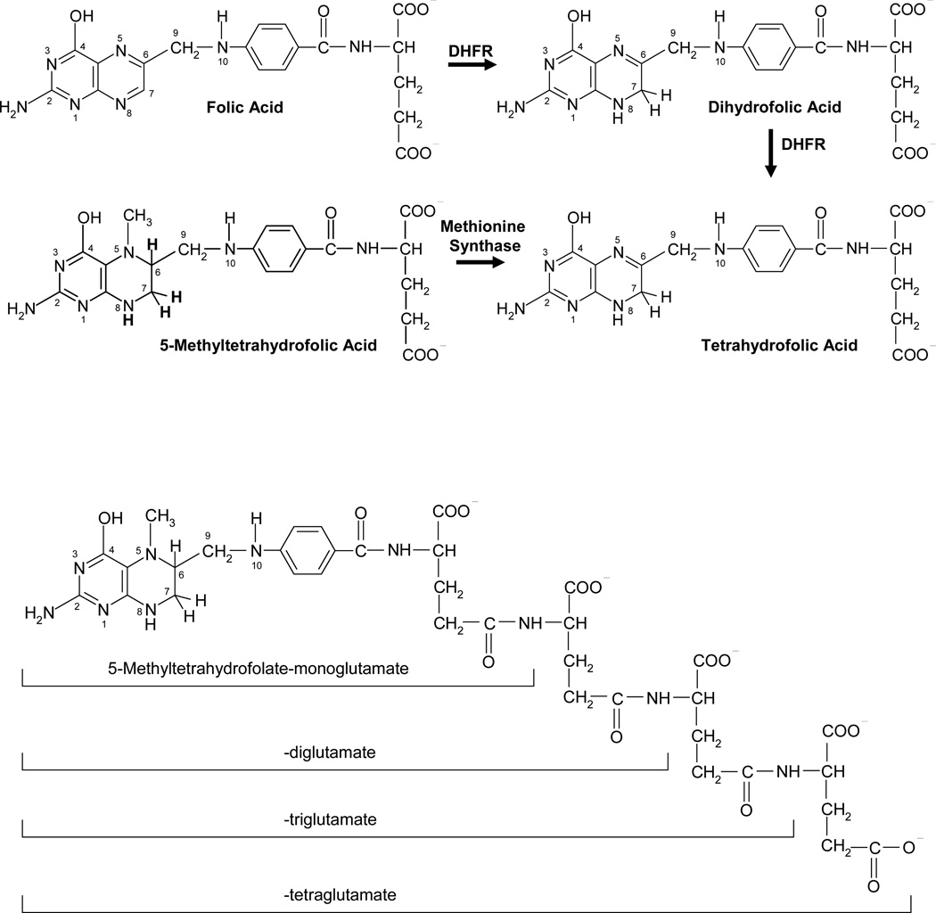
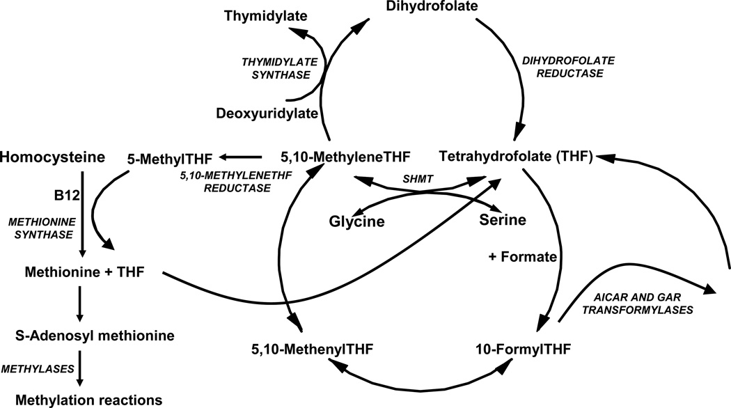
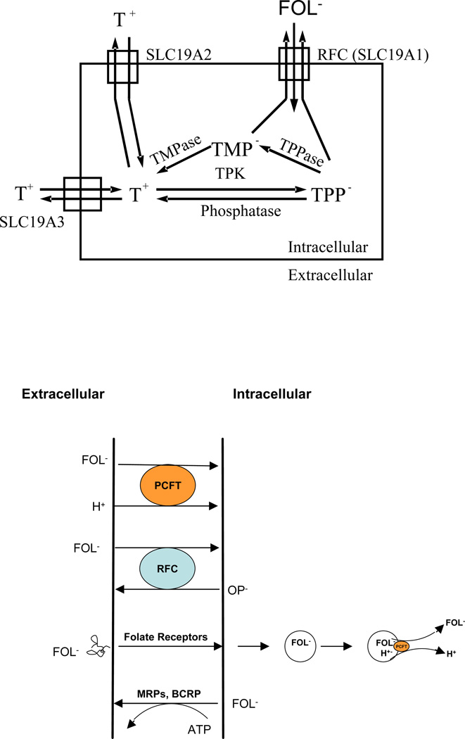
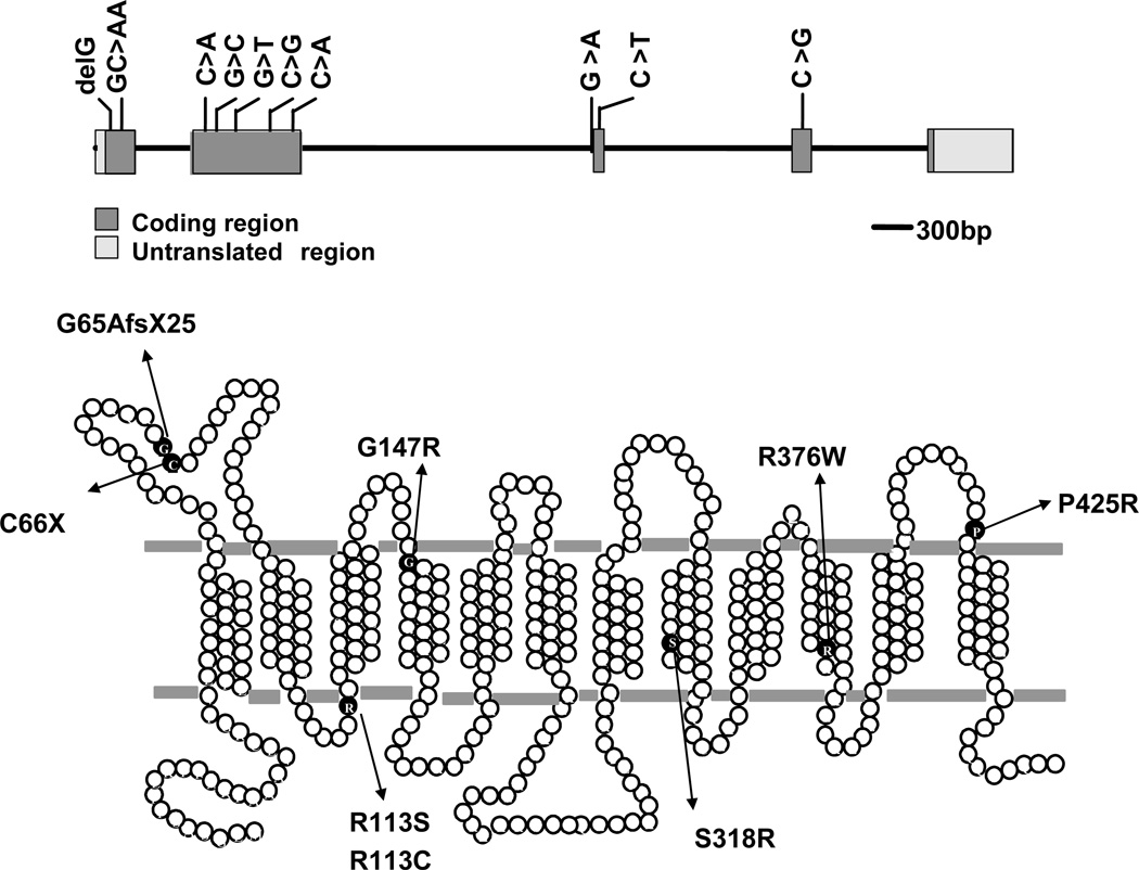
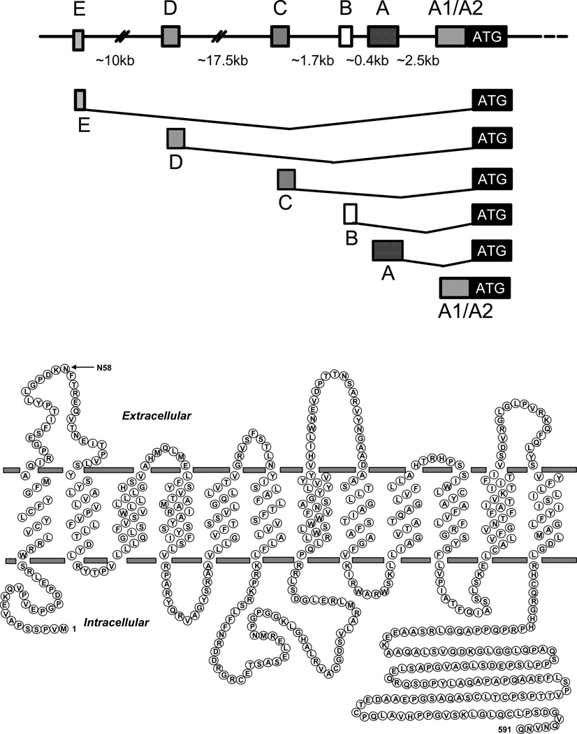
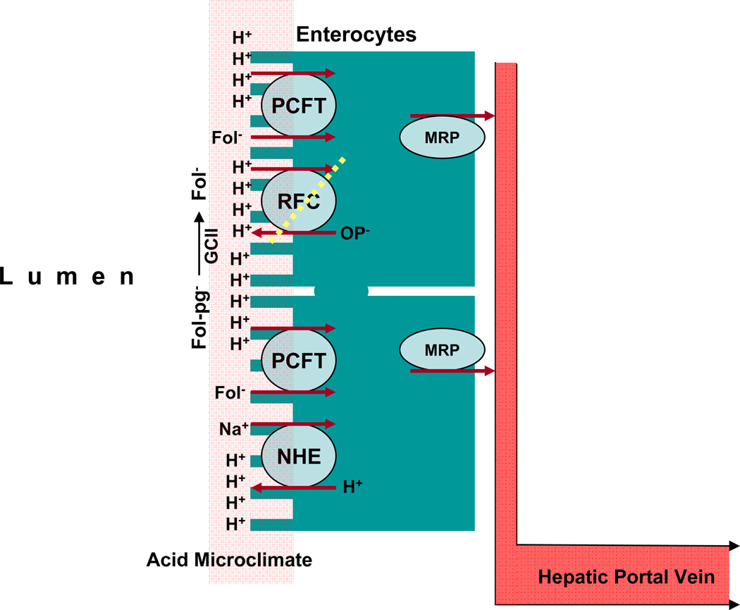
Members of the family of B9 vitamins are commonly known as folates. They are derived entirely from dietary sources and are key one-carbon donors required for de novo nucleotide and methionine synthesis. These highly hydrophilic molecules use several genetically distinct and functionally diverse transport systems to enter cells: the reduced folate carrier, the proton-coupled folate transporter and the folate receptors. Each plays a unique role in mediating folate transport across epithelia and into systemic tissues. The mechanism of intestinal folate absorption was recently uncovered, revealing the genetic basis for the autosomal recessive disorder hereditary folate malabsorption, which results from loss-of-function mutations in the proton-coupled folate transporter gene. It is therefore now possible to piece together how these folate transporters contribute, both individually and collectively, to folate homeostasis in humans. This review focuses on the physiological roles of the major folate transporters, with a brief consideration of their impact on the pharmacological activities of antifolates.


Figures

Figure 1
Figure 1
Upper Panel: Structures of representative folate compounds. Folic acid is reduced to dihydrofolic acid at the positions 7,8 of the B ring which, in turn, is reduced to tetrahydrofolic acid at the positions 5,6 of the B ring in reactions mediated by dihydrofolate reductase (DHFR). Endogenous folic acid is not found in cells. Rather, its presence is related to the consumption of folic acid added to cereals and grains and vitamins. The major oxidized folate in cells is dihydrofolic acid. 5-Methyltetrahydrofolic acid (5-methylTHF) is the major dietary form and the major folate found in blood. It is formed endogenously by the reduction of 5,10-methylene-tetrahydrofolic acid (5,10-methyleneTHF). With the utilization of 5-methylTHF in methionine synthesis, tetrahydrofolate (THF) is generated. These relationships are depicted in Figure 2. The two carboxyl groups of the glutamate moiety are fully ionized at physiological pH. Lower Panel: Structure of 5-methylTHF tetraglutamate. This is one of the 5-methylTHF forms found in nature and in mammalian cells. This is a poly-anion and a very poor substrate for folate transporters and multidrug resistance associated proteins.
Figure 2
Figure 2
Folate metabolic pathways. 5-MethylTHF enters the metabolic cycle following transport into cells where, with homocysteine, it is utilized in the synthesis of methionine mediated by methionine synthase and vitamin B12. The THF product then acquires a carbon from formate or serine, at the N5, N10, or shared between the N5 and N10 positions that, through a series of interconversions, results in several other folate forms that provide carbons for purine, thymidylate, and methionine synthesis. Abbreviations: (AICAR, 5-amino-4-imidazolecarboxamide ribonucleotide; GAR, β-glycinamideribonucleotide; SHMT, serinehydroxymethyltransferase.
Figure 3
Figure 3
Upper panel: Interactions among thiamine, its phosphorylated derivatives, folates, and the SLC19 family of transporters. Thiamine (T+), a cation, is transported into cells via SLC19A2 and SLC19A3. Within cells thiamine is metabolized to thiamine pyrophosphate (TPP), an anion, in a reaction mediated by thiamine pyrophosphate kinase (TPK). TPP can be hydrolyzed to thiamine monophosphate (TMP), also an anion, mediated by thiamine pyrophosphatase (TPPase). Both are substrates for the reduced folate carrier (RFC-SLC19A1) and are exported by that mechanism. Folates enter cells via RFC and are not substrates for the thiamine transporters. Likewise, thiamine is not a substrate for RFC. Lower panel: A schema of the current known folate transporters. The proton-coupled folate transporter (PCFT) is a folate (FOL)-H+ symporter that functions most efficiently in an acidic extracellular environment. The reduced folate carrier (RFC) is an anion exchanger that utilizes the transmembrane organic phosphate (OP) gradient to achieve uphill transport into cells. Folate receptors (FRs) α and β are high- affinity folate binding proteins that transport folates into cells via an endocytic mechanism. Once in the cytoplasm, the vesicle acidifies, folate is release from receptor and is exported from the endosome via PCFT. Folate monoglutamates are exported from cells via the multidrug-associated resistance proteins (MRPs) and the breast cancer resistant protein (BCRP).
Figure 4
Figure 4
Upstream RFC gene structure and membrane topology. Upper Panel: A schematic of the upstream region of the human RFC gene including up to 6 alternate non-coding regions, each preceded by a separate promoter, spanning ∼35 kb upstream of the major translational start site (shown). The alternate promoters transcribe unique RFC transcripts each with a distinct 5’ untranslated region (5’UTRs) fused to a common coding sequence. Alternate splicing for the A1/A2, A, B, and D 5’UTRs have been described. For the A1/A2 and A 5’UTRs, upstream AUGs occur in-frame within the RFC non-coding sequence and result in N-terminally modified hRFC protein isoforms, with 64 and 22 additional N-terminal amino acids, respectively. Lower Panel: A schematic is shown for the membrane topology of the human RFC including 12 transmembrane domains (TMDs), internally oriented N- and C-termini, the large cytosolic loop between TMDs 6 and 7, and the N-glycosylation site at Asparagine 58.
Figure 5
Figure 5
The genomic organization and predicted secondary structure of PCFT and the spectrum of reported mutations in the protein derived from patients with the autosomal recessive disorder, hereditary folate malabsorption (HFM). Upper Panel: The genomic organization of PCFT; the positions of the various base mutations in the first through fourth exons are indicated. Lower Panel: A schematic of a predicted human PCFT topology showing confirmed N-glycosylation sites between the first and second TMDs with the N- and C- termini oriented to the cytoplasm. The location and identity of the mutated amino acid residues involved with HFM are indicated.
Figure 6
Figure 6
Schematic of the sequence of events that occur in the proximal jejunum during the process of hydrolysis and absorption of dietary folate polyglutamates. Dietary 5-methylTHF polyglutamates (Fol-pg) are hydrolyzed to monoglutamate by glutamate carboxypeptidase II (GCII) and then transported into cells via PCFT; both reactions have a low pH optimum and are favored by the low pH within the microenvironment at the jejunal villi (indicated in pink) that is generated by NHE Na+/H+ exchangers. Cellular folates exit the basolateral membrane by an, as yet, unconfirmed mechanism likely related to a member(s) of the multidrug-associated protein (MRP) family of ABC cassette exporters.

Similar articles

Cited by

References

Reference List

    1. Stokstad ELR. Historical perspective on key advances in the biochemistry and physiology of folates. In: Picciano MF, E. Stokstad LR, editors. Folic Acid Metabolism in Health and Disease. New York: Wiley-Liss; 1990. pp. 1–21.
    1. Jacques PF, et al. The effect of folic acid fortification on plasma folate and total homocysteine concentrations. N Engl J Med. 1999;340:1449–1454. - PubMed
    1. Matherly LH, Goldman DI. Membrane transport of folates. Vitam Horm. 2003;66:403–456. - PubMed
    1. Matherly LH, Hou Z, Deng Y. Human reduced folate carrier: translation of basic biology to cancer etiology and therapy. Cancer Metastasis Rev. 2007;26:111–128. - PubMed
    1. Kamen BA, Smith AK. A review of folate receptor alpha cycling and 5-methyltetrahydrofolate accumulation with an emphasis on cell models in vitro. Adv Drug Deliv Rev. 2004;56:1085–1097. - PubMed

9.0 Website

    1. For an in-depth up-to-date review of hereditary folate Malabsorption from the clinical and genetic perspectives. http://www.ncbi.nlm.nih.gov/bookshelf/br.fcgi?book=gene&part=folate-mal.

Publication types

MeSH terms