. 2009 Feb 10;106(6):1903-8.
doi: 10.1073/pnas.0811556106. Epub 2009 Jan 27.

Heterogeneity of natural Foxp3+ T cells: a committed regulatory T-cell lineage and an uncommitted minor population retaining plasticity

Affiliations

Heterogeneity of natural Foxp3+ T cells: a committed regulatory T-cell lineage and an uncommitted minor population retaining plasticity

Noriko Komatsu et al. Proc Natl Acad Sci U S A. .

Abstract

Natural regulatory T cells (T(reg)) represent a distinct lineage of T lymphocytes committed to suppressive functions, and expression of the transcription factor Foxp3 is thought to identify this lineage specifically. Here we report that, whereas the majority of natural CD4(+)Foxp3(+) T cells maintain stable Foxp3 expression after adoptive transfer to lymphopenic or lymphoreplete recipients, a minor fraction enriched within the CD25(-) subset actually lose it. Some of those Foxp3(-) T cells adopt effector helper T cell (T(h)) functions, whereas some retain "memory" of previous Foxp3 expression, reacquiring Foxp3 upon activation. This minority "unstable" population exhibits flexible responses to cytokine signals, relying on transforming growth factor-beta to maintain Foxp3 expression and responding to other cytokines by differentiating into effector T(h) in vitro. In contrast, CD4(+)Foxp3(+)CD25(high) T cells are resistant to such conversion to effector T(h) even after many rounds of cell division. These results demonstrate that natural Foxp3(+) T cells are a heterogeneous population consisting of a committed T(reg) lineage and an uncommitted subpopulation with developmental plasticity.


Conflict of interest statement

The authors declare no conflict of interest.

Figures

Fig. 1.
Fig. 1.
A fraction of CD4+Foxp3+ T cells lose Foxp3 expression in lymphopenic and normal lymphoreplete conditions. (A) CD4+EGFP+ and EGFP T cells were sorted from Foxp3EGFP Ly5.2 and Foxp3EGFP Ly5.1 mice, respectively, and adoptively transferred into RAG2−/− mice, either alone (1 × 10⁵ each) or mixed at a 1:1 or 1:10 ratio (1 × 10⁵ EGFP+ plus 1 × 10⁵ or 1 × 10⁶ EGFP). Four weeks after transfer, LN cells were stained for CD4, TCRβ and Ly5.1. Shown are representative EGFP expression profiles of the Foxp3+ (Ly5.1) or Foxp3 (Ly5.1+) donor-derived CD4+TCRβ+ cells recovered from the indicated host mice. (B) 1 × 10⁵ CD4+EGFP+ T cells sorted from Foxp3EGFP Ly5.1/Thy1.2 mice were adoptively transferred into Ly5.2/Thy1.1 host mice 4 and 8 weeks earlier. Pooled LN and spleen cells were first enriched for donor T cells by depleting Thy1.1+, Ig+, and adherent cells by panning, and were stained for Ly5.1, Thy1.2, and CD4. A representative EGFP and CD4 profile of the Ly5.1+Thy1.2+ donor cells (left) and frequencies of EGFP cells in CD4+Ly5.1+Thy1.2+ donor T cells (right) are shown. Each symbol indicates individual host mouse.
Fig. 2.
Fig. 2.
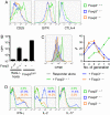
CD4+Foxp3+ T cells lose Treg phenotype and produce inflammatory cytokines upon down-regulation of Foxp3. (A) CD4+EGFP+ or EGFP T cells were transferred into RAG2−/− mice as in Fig. 1A. Four weeks later, LN cells were stained for TCRβ, CD4, and the indicated markers. Shown are representative profiles of CD25, GITR, and CTLA-4 expression on TCRβ+CD4+EGFP+ (Foxp3+→+) and EGFP (Foxp3+→−) cells from the recipients of EGFP+ cells and on TCRβ+CD4+EGFP cells from the recipients of EGFP cells (Foxp3−→−). (B–D) CD4+EGFP+ or EGFP T cells from Foxp3EGFP Ly5.1 mice were transferred into RAG2−/− (B, C) or CD3ε−/− (D) mice 4 weeks earlier. Pooled LN and spleen cells were stained for CD4, and Ly5.1, and Foxp3+→+, Foxp3+→−, and Foxp3−→− CD4+Ly5.1+ donor cells were sorted. (B) Indicated populations were stimulated in vitro with anti-CD3, and proliferation was measured by [3H]thymidine incorporation. (C) CFSE-labeled CD4+CD25 responder T cells from B6 (Ly5.2) mice were stimulated in vitro either alone or together with an equal number of the indicated populations. On day 3, cells were stained for Ly5.1 and CD4, and CFSE dilution on the CD4+Ly5.1 responder population was determined. Shown are representative CFSE profiles of responder T cells from each group (left) and frequencies of cells of the indicated generation (right). (D) Indicated populations were stimulated with PMA and ionomycin and production of IFN-γ, IL-2, and IL-17 was assessed by intracellular staining.
Fig. 3.
Fig. 3.
Preferential re-induction of Foxp3 expression in Foxp3down-regulated T-cells in vitro and in vivo. CD4+Foxp3+→− and Foxp3−→− donor T cells were sorted from CD3ε−/− hosts as described in Fig. 2B-D. (A) Sorted cells were stimulated in vitro with anti-CD3→CD28 beads, and analyzed for CD4 and EGFP expression on day 3. (B) Foxp3+→− T cells were stimulated as in A with or without a mixture of anti-TGF-β mAbs and TGFβRII-Fc. (C) 1 × 10⁵ Foxp3+→− or Foxp3−→− T cells or EGFP T cells from Foxp3EGFP mice were transferred into new CD3ε−/− host mice. Four weeks after the transfer, LN and spleen (not depicted) cells were stained for CD4 and Ly5.1, and analyzed for EGFP expression. Shown are frequencies of (re-)induced Foxp3+ T cells in CD4+Ly5.1+ donor T cells. Each symbol represents individual host mouse.
Fig. 4.
Fig. 4.
Foxp3-down-regulated T-cells have proliferated extensively in lymphopenic mice. CD4+hCD2+ T cells (4 × 10⁵) from Foxp3hCD2 Ly5.1 mice were labeled with CFSE and injected into RAG2−/− mice. On the indicated time points, LN and spleen cells from all recipient mice (n = 3) were pooled and stained for hCD2, Ly5.1, and Ly5.2. Shown are hCD2 and CFSE profiles on Ly5.1+Ly5.2 donor cells.
Fig. 5.
Fig. 5.
CD4+Foxp3+CD25 T cells exhibit lower and less stable Foxp3 expression than CD4+Foxp3+CD25 high T cells. (A) LN cells from Foxp3hCD2 mice were stained for hCD2, CD4, and CD25. Representative CD25 and hCD2 expression profiles on CD4+ cells are shown. The number above each gate indicates the mean fluorescent intensity of hCD2. (B) Sorted CD4+hCD2+CD25 and CD25high T cells were stimulated in vitro with anti-CD3/CD28 beads in the presence of IL-2 and analyzed for hCD2 and CD4 expression on day 3. (C) 1 × 10⁵ Ly5.1 CD4+hCD2+CD25 and CD25high T cells were transferred into RAG2−/− mice, and LN cells were stained for hCD2, Ly5.1, and Ly5.2 on day 5. Shown are representative hCD2 and Ly5.1 expression profiles (left) and frequencies of hCD2 cells (right) in donor (Ly5.1+Ly5.2) cells. (D) Ly5.1 CD4+hCD2+CD25 and CD25high T cells were stained with CFSE and transferred into Foxp3hCD2 Ly5.2 mice (4 × 10⁵ cells). On day 14, pooled LN and spleen cells were stained for Ly5.1, Ly5.2, and hCD2 and enriched for donor Ly5.1+ cells by magnetic sorting before analysis. Representative CFSE and hCD2 profiles on Ly5.1+Ly5.2 cells are shown.
Fig. 6.
Fig. 6.
Only CD4+Foxp3+CD25 T cells require TGF-β for Foxp3 maintenance. These cells can be instructed by cytokines to differentiate into effector Th, whereas CD4+Foxp3+CD25high T cells cannot. (A, B) Sorted CD4+hCD2+CD25 and CD25high T cells were stimulated in vitro with anti-CD3/CD28 beads in the presence of IL-2 with or without the indicated cytokines or mAbs. On day 3, cells were stained with anti-hCD2 or its isotype control mAbs. (C) Sorted CD4+hCD2+ and hCD2 T cells were stimulated in vitro with anti-CD3/CD28 beads in the presence of IL-4 or IL-6. On day 5, cells were re-stimulated and stained for IL-4 or IL-17 production. (D) Total Ly5.1 CD4+Foxp3hCD2+ T cells were transferred into CD3ε−/− mice 4 weeks earlier. Sorted CD4+Foxp3+→+ Ly5.1+ and hCD2+ T cells from Foxp3hCD2 mice were stimulated in the presence of the indicated reagents.

References

    1. Sakaguchi S. Naturally arising CD4+ regulatory T cells for immunologic self-tolerance and negative control of immune responses. Annu Rev Immunol. 2004;22:531–562. - PubMed
    1. Hori S, Nomura T, Sakaguchi S. Control of regulatory T-cell development by the transcription factor Foxp3. Science. 2003;299:1057–1061. - PubMed
    1. Fontenot JD, Gavin MA, Rudensky AY. Foxp3 programs the development and function of CD4+CD25+ regulatory T cells. Nat Immunol. 2003;4:330–336. - PubMed
    1. Khattri R, Cox T, Yasayko SA, Ramsdell F. An essential role for Scurfin in CD4+CD25+ T regulatory cells. Nat Immunol. 2003;4:337–342. - PubMed
    1. Fontenot JD, et al. Regulatory T-cell lineage specification by the forkhead transcription factor Foxp3. Immunity. 2005;22:329–341. - PubMed

Publication types

MeSH terms

LinkOut - more resources