. 2009 Apr;37(6):1778-88.
doi: 10.1093/nar/gkp015. Epub 2009 Jan 27.

Improved tetracycline repressors for gene silencing in mycobacteria

Affiliations

Improved tetracycline repressors for gene silencing in mycobacteria

Marcus Klotzsche et al. Nucleic Acids Res. 2009 Apr.

Abstract

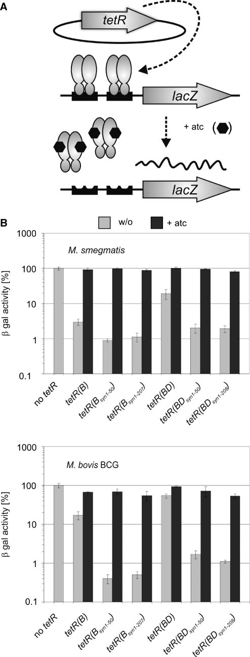
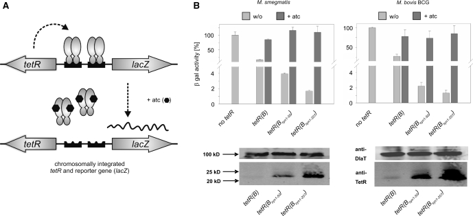
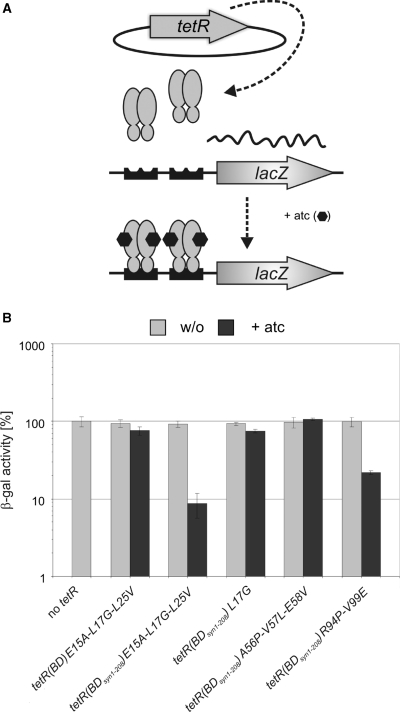
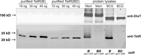
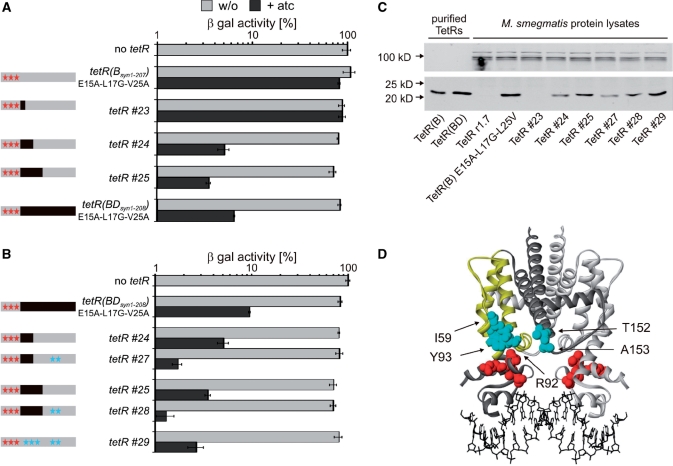
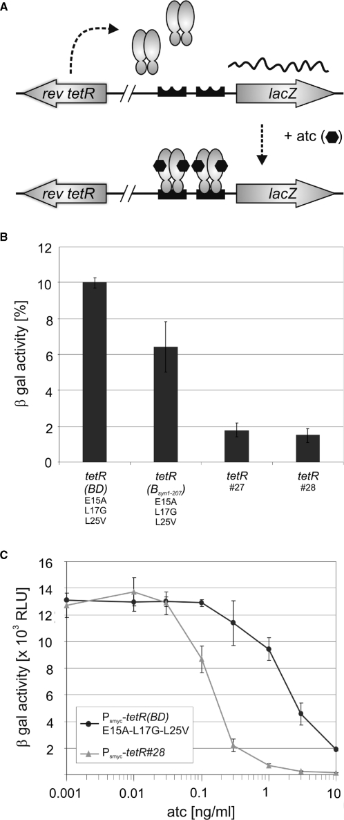
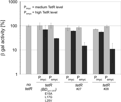
Tetracycline repressor (TetR)-controlled expression systems have recently been developed for mycobacteria and proven useful for the construction of conditional knockdown mutants and their analysis in vitro and during infections. However, even though these systems allowed tight regulation of some mycobacterial genes, they only showed limited or no phenotypic regulation for others. By adapting their codon usage to that of the Mycobacterium tuberculosis genome, we created tetR genes that mediate up to approximately 50-fold better repression of reporter gene activities in Mycobacterium smegmatis and Mycobacterium bovis BCG. In addition to these repressors, for which anhydrotetracycline (atc) functions as an inducer of gene expression, we used codon-usage adaption and structure-based design to develop improved reverse TetRs, for which atc functions as a corepressor. The previously described reverse repressor TetR only functioned when expressed from a strong promoter on a multicopy plasmid. The new reverse TetRs silence target genes more efficiently and allowed complete phenotypic silencing of M. smegmatis secA1 with chromosomally integrated tetR genes.


Figures

Figure 1.
Figure 1.
Impact of tetR codon usage adaptation on repression of chromosomally encoded β-galactosidase activities by episomally encoded wt TetRs. (A) Genetic organization of the assay strains. The Pmyc1tetO-lacZ reporter gene cassette was integrated into the mycobacteriophage L5 attachment site. The Pimyc-tetR genes were located on episomally replicating plasmids. Binding of TetR to tetOs in the absence of atc caused repression of Pmyc1tetO. Symbols: TetRs, gray ovals; tetOs, black boxes; atc, black hexagons. (B) β-Galactosidase activities (β-gal). Expressed TetR variants are identified underneath the graph. Values were normalized to the β-galactosidase activity measured in the absence of TetR, which was set to 100%. Bars represent averages of three measurements and are representative of at least two independent experiments. Error bars indicate standard deviations.
Figure 2.
Figure 2.
Impact of tetR codon usage adaptation on repression of chromosomally encoded β-galactosidase activities by chromosomally encoded wt TetRs. (A) Genetic organization of the assay strains. Plasmids containing both, the Pmyc1tetO-lacZ reporter gene cassette and the Psmyc-tetR genes were integrated into the chromosome via the L5 attachement site. Symbols: TetRs, gray ovals; tetOs, black boxes; atc, black hexagons. (B) β-galactosidase activities (β-gal) and steady-state TetR protein levels detected by a monoclonal anti-TetR antibody. Expressed TetR variants are identified underneath the bar graph. Values were normalized to the β-galactosidase activity measured in the absence of TetR, which was set to 100%. Bars represent averages of three measurements and are representative of at least two independent experiments. Error bars indicate standard deviations. Western blots are shown below the respective bar graphs. The dihydrolipoamide acyltransferase (DlaT) signal was used as a loading control and detected by a polyclonal anti-DlaT antibody.
Figure 3.
Figure 3.
Impact of tetR codon usage adaptation on repression of chromosomally encoded β-galactosidase activities by episomally encoded reverse TetRs. (A) Genetic organization of the assay strains. The Pmyc1tetO-lacZ reporter gene cassette was integrated into the mycobacteriophage L5 attachment site. The Pimyc-tetR genes were located on episomally replicating plasmids. Binding of revTetR to tetOs in the presence of atc caused repression of Pmyc1tetO. Symbols: TetRs, gray ovals; tetOs, black boxes; atc, black hexagons. (B) β-galactosidase activities (β-gal). Expressed TetR variants are identified underneath the bar graph. Values were normalized to the β-galactosidase activity measured in the absence of TetR, which was set to 100%. Bars represent averages of three measurements and are representative of at least two independent experiments. Error bars indicate standard deviations.
Figure 4.
Figure 4.
Comparison of TetR steady state levels in M. smegmatis and M. bovis BCG containing tetR(B)syn1–207 or tetR(BD)syn1–208. Protein lysates were from M. smegmatis (labeled ‘Msm’) and M. bovis BCG (labeled ‘BCG’) containing episomally replicating Pimyc-tetR plasmids. A monoclonal anti-TetR antibody recognizing an epitope within the DNA-binding region of TetR was used to detect purified TetR as well as TetR in protein lysates from M. smegmatis and M. bovis BCG. The DlaT signal was used as a loading control and detected by a polyclonal anti-DlaT antibody.
Figure 5.
Figure 5.
Analysis of reverse TetR chimeras in M. smegmatis. (A) β-galactosidase activities (β-gal). Expressed TetR variants are specified left of the bar graph. In the schemas on the left, gray and black rectangles indicate sequences derived from tetR(B) and tetR(D) respectively. Red stars indicate the location of mutation causing the reverse phenotype of TetR r1.7. Blue stars indicate TetR(B) to TetR(D) mutations. β-Gal values were normalized to the β-galactosidase activity measured in the absence of TetR, which was set to 100%. Bars represent averages of three measurements and are representative of at least two independent experiments. Error bars indicate standard deviations. (B) β-galactosidase activities (β-gal). As described for (A). (C) Western blots. Protein lysates were prepared from M. smegmatis expressing TetR variants encoded by episomally replicating plasmids. TetR was detected by a monoclonal anti-TetR antibody (lower panel) and the dihydrolipoamide acyltransferase (DlaT) signal was used as a loading control and detected by a polyclonal anti-DlaT antibody (upper panel). (D) Structure of tetO-bound TetR(D) (22). The two monomers are shown in light and dark gray. The side chains of the amino acids that are mutated in and cause the reverse phenotype of TetR r1.7 are shown in red. In the dark gray monomer helices 4 to 7 are drawn in yellow. Side chains that are different in TetR(B) and TetR(D) and within a radius of 15 Å to positions 15, 17 and 25 are shown in light blue (for clarity sake only within one monomer).
Figure 6.
Figure 6.
Repression of lacZ by chromosomally encoded reverse TetRs. (A) Genetic organization of the assay strains. The Pmyc1tetO-lacZ reporter gene cassette was integrated into the mycobacteriophage tweety attachment site. The Psmyc-tetR genes were integrated into the mycobacteriophage L5 attachment site. Symbols: TetRs, gray ovals; tetOs, black boxes; atc, black hexagons. (B) β-galactosidase activities (β-gal) with 300 ng/ml atc. Expressed TetR variants are specified below the bar graph. β-gal values were normalized to the β-galactosidase activity measured in the absence of TetR, which was set to 100%. Bars represent averages of three measurements and are representative of at least two independent experiments. Error bars indicate standard deviations. (C) β-galactosidase activities (β-gal) with different atc concentrations. Black circles and gray triangles represent data from M. smegmatis containing Psmyc-tetR(BD)E15A-L17G-L25V and Psmyc-tetR#28, respectively.
Figure 7.
Figure 7.
Analysis of reverse TetRs in M. bovis BCG. TetR-mediated repression was measured using the chromosomally integrated Pmyc1tetO-lacZ reporter. TetR variants were expressed from episomally replicating plasmids. β-galactosidase activities were analyzed in the absence (light gray bars) and presence of 300 ng/ml atc (dark gray bars) using a fluorescent-based assay as explained in ‘Materials and methods’ sections. Obtained values were normalized to the β-galactosidase activity measured in the absence of TetR, which was set to 100%. Bars represent averages of three measurements and are representative of at least two independent experiments. Error bars indicate standard deviations.

References

    1. Cole ST, Brosch R, Parkhill J, Garnier T, Churcher C, Harris D, Gordon SV, Eiglmeier K, Gas S, Barry CE, III, et al. Deciphering the biology of Mycobacterium tuberculosis from the complete genome sequence. Nature. 1998;393:537–544. - PubMed
    1. Sassetti CM, Boyd DH, Rubin EJ. Genes required for mycobacterial growth defined by high density mutagenesis. Mol. Microbiol. 2003;48:77–84. - PubMed
    1. Chalut C, Botella L, de Sousa-D’Auria C, Houssin C, Guilhot C. The nonredundant roles of two 4′-phosphopantetheinyl transferases in vital processes of Mycobacteria. Proc. Natl Acad. Sci. USA. 2006;103:8511–8516. - PMC - PubMed
    1. Ehrt S, Guo XV, Hickey CM, Ryou M, Monteleone M, Riley LW, Schnappinger D. Controlling gene expression in mycobacteria with anhydrotetracycline and Tet repressor. Nucleic Acids Res. 2005;33:e21. - PMC - PubMed
    1. Gandotra S, Schnappinger D, Monteleone M, Hillen W, Ehrt S. In vivo gene silencing identifies the Mycobacterium tuberculosis proteasome as essential for the bacteria to persist in mice. Nat. Med. 2007;13:1515–1520. - PMC - PubMed

Publication types

Substances