Expression of bile acid transporting proteins in Barrett's esophagus and esophageal adenocarcinoma
- PMID: 19174784
- PMCID: PMC4450811
- DOI: 10.1038/ajg.2008.85
Expression of bile acid transporting proteins in Barrett's esophagus and esophageal adenocarcinoma
Abstract
Objectives: Barrett's esophagus (BE) is a metaplastic lesion characterized by replacement of the normal squamous epithelium by columnar intestinal epithelium containing goblet cells. It is speculated that this process is an adaptation to protect cells from components of refluxate, such as gastric acid and bile acids. In contrast to the normal squamous epithelium, enterocytes of the distal ileum are adapted to transport bile acids from the intestinal lumen. Several bile acid transporters are utilized for effective removal of bile acids, including the apical sodium-dependent bile acid transporter (ASBT), the ileal bile acid-binding protein (IBABP), and the multidrug-resistant protein 3 (MRP3). We hypothesized that one of the possible functions of newly arising metaplastic epithelium, in the esophagus, is to transport bile acids. Our major goal was to evaluate the expression of bile acid transporters in normal squamous epithelium, BE with different grades of dysplasia, and esophageal adenocarcinoma (EAC).
Methods: A total of 101 patients were included in this study. Immunohistochemistry (IHC) and reverse transcriptase (RT)-PCR were used to detect the expression of these transporters at the mRNA and protein levels.
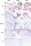
Results: Our immunohistochemical studies showed that all three bile acid transporters are expressed in BE glands, but not in squamous epithelium. ASBT was found in the apical border in BE biopsies. The highest frequency of ASBT expression was in patients with nondysplastic BE (9 of 15, 60%), and a progressive loss of ASBT was observed through the stages of dysplasia. ASBT was not detected in EAC (0 of 15). IBABP staining was observed in the cytoplasm of BE epithelial surface cells. Expression of IBABP was found in 100% of nondysplastic BE (14 of 14), in 93% of low-grade dysplasia (LGD, 15 of 16), in 73% of high-grade dysplasia (HGD, 10 of 14), and in 33% of EAC (5 of 15). MRP3 was expressed in the basolateral membrane in 93% of nondysplastic BE (13 of 14), in 60% of LGD (10 of 16), and in 86% of HGD (11 of 13). Only weak MRP3 staining was detected in EAC biopsies (5 of 15, 33%). In addition, RT-PCR studies showed increased expression of mRNA coding for ASBT (6.1x), IBABP (9.1x), and MRP3 (2.4x) in BE (N=13) compared with normal squamous epithelium (N=15). Significantly increased mRNA levels of IBABP (10.1x) and MRP3 (2.5x) were also detected in EAC (N=21) compared with normal squamous epithelium.
Conclusions: We found that bile acid transporters expression is increased in BE tissue at the mRNA and protein levels and that expression of bile acid transporter proteins decreased with progression to cancer.
Figures


References
-
- Ronkainen J, Aro P, Storskrubb T, et al. Prevalence of Barrett’s esophagus in the general population: an endoscopic study. Gastroenterology. 2005;129:1825–31. - PubMed
-
- Sampliner RE. Epidemiology, pathophysiology, and treatment of Barrett’s esophagus: reducing mortality from esophageal adenocarcinoma. Med Clin North Am. 2005;89:293–312. - PubMed
Publication types
MeSH terms
Substances
Grants and funding
LinkOut - more resources
Full Text Sources
Medical

