

Automatic segmentation of the facial nerve and chorda tympani in CT images using spatially dependent feature values
- PMID: 19175097
- PMCID: PMC2673604
- DOI: 10.1118/1.3005479
Automatic segmentation of the facial nerve and chorda tympani in CT images using spatially dependent feature values
Abstract
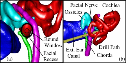
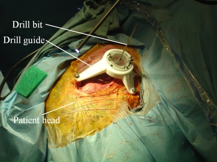
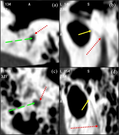
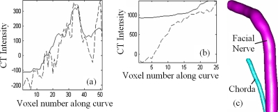
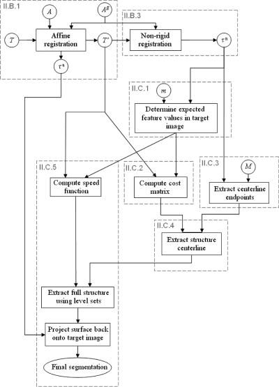
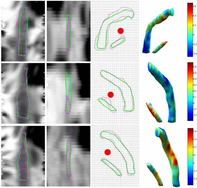
In cochlear implant surgery, an electrode array is permanently implanted in the cochlea to stimulate the auditory nerve and allow deaf people to hear. A minimally invasive surgical technique has recently been proposed-percutaneous cochlear access-in which a single hole is drilled from the skull surface to the cochlea. For the method to be feasible, a safe and effective drilling trajectory must be determined using a preoperative CT. Segmentation of the structures of the ear would improve trajectory planning safety and efficiency and enable the possibility of automated planning. Two important structures of the ear, the facial nerve and the chorda tympani, are difficult to segment with traditional methods because of their size (diameters as small as 1.0 and 0.3 mm, respectively), the lack of contrast with adjacent structures, and large interpatient variations. A multipart, model-based segmentation algorithm is presented in this article that accomplishes automatic segmentation of the facial nerve and chorda tympani. Segmentation results are presented for ten test ears and are compared to manually segmented surfaces. The results show that the maximum error in structure wall localization is approximately 2 voxels for the facial nerve and the chorda, demonstrating that the method the authors propose is robust and accurate.
Figures

Similar articles
-
Automatic determination of optimal linear drilling trajectories for cochlear access accounting for drill-positioning error.Int J Med Robot. 2010 Sep;6(3):281-90. doi: 10.1002/rcs.330. Int J Med Robot. 2010. PMID: 20812268 Free PMC article.
-
Automatic segmentation of the facial nerve and chorda tympani in pediatric CT scans.Med Phys. 2011 Oct;38(10):5590-600. doi: 10.1118/1.3634048. Med Phys. 2011. PMID: 21992377 Free PMC article.
-
Automatic segmentation of intra-cochlear anatomy in post-implantation CT of unilateral cochlear implant recipients.Med Image Anal. 2014 Apr;18(3):605-15. doi: 10.1016/j.media.2014.02.001. Epub 2014 Feb 18. Med Image Anal. 2014. PMID: 24650801 Free PMC article.
-
[Surgical technique in cochlear implantation].HNO. 2009 Jul;57(7):663-70. doi: 10.1007/s00106-009-1948-6. HNO. 2009. PMID: 19554272 Review. German.
-
Cochlear implantation in children with anomalous cochleovestibular anatomy.Laryngoscope. 2005 Jan;115(1 Pt 2 Suppl 106):1-26. doi: 10.1097/00005537-200501001-00001. Laryngoscope. 2005. PMID: 15626926 Review.
Cited by
-
A Compact, Bone-Attached Robot for Mastoidectomy.J Med Device. 2015 Sep;9(3):0310031-310037. doi: 10.1115/1.4030083. J Med Device. 2015. PMID: 26336572 Free PMC article.
-
Validation of minimally invasive, image-guided cochlear implantation using Advanced Bionics, Cochlear, and Medel electrodes in a cadaver model.Int J Comput Assist Radiol Surg. 2013 Nov;8(6):989-95. doi: 10.1007/s11548-013-0842-6. Epub 2013 Apr 30. Int J Comput Assist Radiol Surg. 2013. PMID: 23633113 Free PMC article.
-
A manually operated, advance off-stylet insertion tool for minimally invasive cochlear implantation surgery.IEEE Trans Biomed Eng. 2012 Oct;59(10):2792-800. doi: 10.1109/TBME.2012.2210220. Epub 2012 Jul 25. IEEE Trans Biomed Eng. 2012. PMID: 22851233 Free PMC article.
-
Automatic determination of optimal linear drilling trajectories for cochlear access accounting for drill-positioning error.Int J Med Robot. 2010 Sep;6(3):281-90. doi: 10.1002/rcs.330. Int J Med Robot. 2010. PMID: 20812268 Free PMC article.
-
Deep learning-based approach for the automatic segmentation of adult and pediatric temporal bone computed tomography images.Quant Imaging Med Surg. 2023 Mar 1;13(3):1577-1591. doi: 10.21037/qims-22-658. Epub 2023 Feb 10. Quant Imaging Med Surg. 2023. PMID: 36915310 Free PMC article.
References
-
- Labadie R. F., Choudhury P., Cetinkaya E., Balachandran R., Haynes D. S., Fenlon M., Juscyzk S., and Fitzpatrick J. M., “Minimally-invasive, image-guided, facial-recess approach to the middle ear: Demonstration of the concept of percutaneous cochlear access in-vitro,” Otol. Neurotol. ONTEAE 26, 557–562 (2005). - PubMed
-
- Fitzpatrick J. M., Konrad P. E., Nickele C., Cetinkaya E., and Kao C., “Accuracy of customized miniature stereotactic platforms,” Stereotact. Funct. Neurosurg. ZZZZZZ 83, 25–31 (2005). - PubMed
-
- Noble J. H., Warren F. M., Labadie R. F., Dawant B. M., and Fitzpatrick J. M., “Determination of drill paths for percutaneous cochlear access accounting for target positioning error,” Proc. SPIE PSISDG 6509, 650925.1–650925.10 (2007).
Publication types
MeSH terms
Grants and funding
LinkOut - more resources
Full Text Sources
Other Literature Sources
Medical
