. 2009 Feb;29(3):447-54.
doi: 10.1111/j.1460-9568.2008.06591.x. Epub 2009 Jan 17.

Progesterone reverses 17beta-estradiol-mediated neuroprotection and BDNF induction in cultured hippocampal slices

Affiliations

Progesterone reverses 17beta-estradiol-mediated neuroprotection and BDNF induction in cultured hippocampal slices

Claudia C Aguirre et al. Eur J Neurosci. 2009 Feb.

Abstract

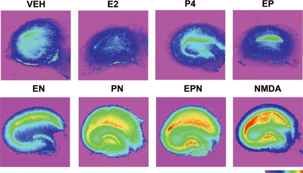
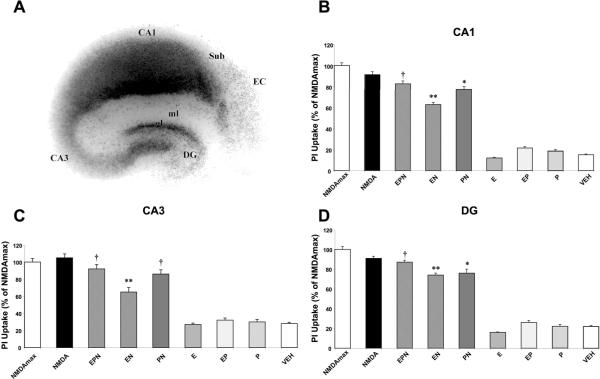
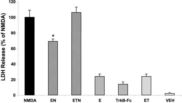
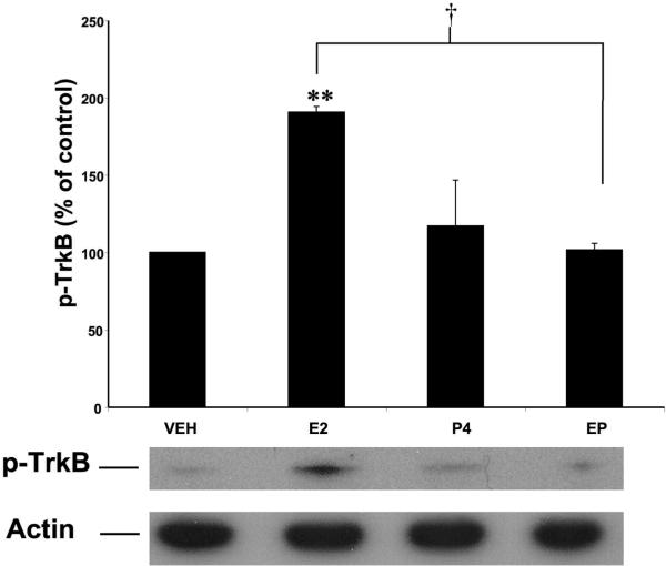
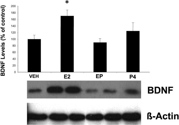
Due to the many similarities in mechanisms of action, targets and effects, progesterone (P4), estrogen and neurotrophins have been implicated in synaptic plasticity as well as in neuroprotection and neurodegeneration. In this study, we examined the interactions between 17beta-estradiol (E2) and P4 and brain-derived neurotrophic factor (BDNF) on both plasticity and excitotoxicity in rat cultured hippocampal slices. First, we evaluated the neuroprotective effects of E2 and P4 against N-methyl-D-aspartate (NMDA) toxicity in cultured rat hippocampal slices. As previously reported, pretreatment with 10 nm E2 (24 h) was neuroprotective against NMDA toxicity. However, P4 (10 nm) added 20 h after E2 treatment for 4 h reversed its protective effect. In addition, the same E2 treatment resulted in an increase in BDNF protein levels as well as in activation of its receptor, TrkB, while addition of P4 attenuated E2-mediated increase in BDNF and TrkB levels. Furthermore, E2-mediated neuroprotection was eliminated by a BDNF scavenger, TrkB-Fc. Our results indicate that E2 neuroprotective effects are mediated through the BDNF pathway and that, under certain conditions, P4 antagonizes the protective effect of estrogen.


Figures

Figure 1
Figure 1. P4-mediated reversal of E2-induced neuroprotection against NMDA toxicity in cultured hippocampal slices
A. Treatment protocol timeline for hormones and NMDA. Treatment with P4 (10 nM), E2 (10 nM), or NMDA (50 μM) was performed as indicated in the diagram. Vehicle was administered for the length of the experiment (not shown). B. LDH release in the medium measured 24 h after NDMA treatment. Medium was collected 24 h after initiation of NMDA treatment, and LDH activity was determined as described under Material and Methods. Results are means ± S.E.M of 7 experiments and are expressed as percent of release in NMDA-treated slices. Statistical significance was analyzed by ANOVA followed by Tukey's test for individual comparisons. *p < 0.001 vs. NMDA; † p < 0.001 vs. E2 + NMDA (EN); NS: not significant.
Figure 2
Figure 2. Effects of E2 and P4 on NMDA-induced neuronal damage in cultured hippocampal slices, assessed with PI uptake
Top Row: Representative images of PI uptake in slices treated with vehicle (VEH), E2 (10 nM), P4 (10 nM) or E2 (10 nM) + P4 (10 nM) (EP). Bottom row: Representative images of PI uptake in slices treated with E2 (10 nM) + NMDA (50 μM) (EN), P4 (10 nM) + NMDA (PN), E2 + P4 + NMDA (EPN) and NMDA alone. NMDA treatment resulted in increased levels of fluorescence intensity, and estrogen attenuated this increase. P4 reversed the protective effects of E2. Color bar represents fluorescence intensity scale, with red illustrating highest intensity.
Figure 3
Figure 3. Quantitative analysis of PI uptake in cultured hippocampal slices
Images of slices treated as in Figure 2 were analyzed as described under Materials and Methods. Regions analyzed (CA1, CA3 and DG) are indicated in A. Results are expressed in percent of fluorescent intensity measured in slices treated with NMDA (50 μM) for 24 h, and are means ± S.E.M. of 12–14 images obtained in 6–8 slices. Statistical significance was analyzed by ANOVA followed by Tukey's test for individual comparisons. ** p < 0.001 as compared to NMDA treatment alone; * p < 0.05 as compared to NMDA treatment alone; † p < 0.05 as compared to treatment with E2 + NMDA (EN).
Figure 4
Figure 4. BDNF mediates E2 neuroprotective effects against NMDA toxicity in cultured hippocampal slices
Cultured hippocampal slices were treated as shown in Figure 1 and TrkB-Fc (0.5 μg/mL) was present during the duration of E2 treatment. Medium was collected 24 h after NMDA treatment and LDH assayed as indicated under Materials and Methods. Results are expressed as percent of LDH release measured in medium of slices treated with NMDA for 3 h and are means ± S.E.M. of 5–6 experiments. Statistical significance was analyzed by ANOVA followed by Tukey's test for individual comparisons. * p < 0.001 as compared to NMDA treated slices.
Figure 5
Figure 5. Effects of E2 and P4 on TrkB activation/phosphorylation in cultured hippocampal slices
Cultured hippocampal slices were treated with E2 (10 nM), P4 (10 nM) or E2 + P4 as described in Figure 1. At the end of treatment, slices were collected and processed for western blot analysis of p-TrkB (bottom). Levels of p-TrkB were corrected with those of β-actin and results were expressed as percent of values found in vehicle-treated slices; they are means ± S.E.M. of 4–5 experiments. Statistical significance was analyzed by ANOVA followed by Tukey's test for individual comparisons. ** p < 0.001 as compared to vehicle-treated slices; † p < 0.001 as compared to E2 treated slices.
Figure 6
Figure 6. Effects of E2 and P4 on BDNF levels in cultured hippocampal slices
Cultured hippocampal slices were treated with E2 (10 nM), P4 (10 nM) or their combination according to the protocol shown in Fig. 1A. Control slices were treated with vehicle. Slices were collected at the end of treatment, and processed for determination of BDNF levels with western blots as described under Material and Methods. Results were normalized to levels of β-actin and were expressed as percent of control; they are means ± S.E.M. of 7 experiments. Statistical significance was analyzed by ANOVA followed by Tukey's test for individual comparisons. * p < 0.05 as compared to control to E2 + P4-treated samples.

References

    1. Alves SE, McEwen BS, Hayashi S, Korach KS, Pfaff DW, Ogawa S. Estrogen-regulated progestin receptors are found in the midbrain raphe but not hippocampus of estrogen receptor alpha (ER alpha) gene-disrupted mice. J Comp Neurol. 2000;427(2):185–195. - PubMed
    1. Bi R, Broutman G, Foy MR, Thompson RF, Baudry M. The tyrosine kinase and mitogen-activated protein kinase pathways mediate multiple effects of estrogen in hippocampus. Proc Natl Acad Sci U S A. 2000;97(7):3602–3607. - PMC - PubMed
    1. Bimonte-Nelson HA, Nelson ME, Granholm AC. Progesterone counteracts estrogen-induced increases in neurotrophins in the aged female rat brain. Neuroreport. 2004;15(17):2659–2663. - PubMed
    1. Blurton-Jones M, Kuan PN, Tuszynski MH. Anatomical evidence for transsynaptic influences of estrogen on brain-derived neurotrophic factor expression. J Comp Neurol. 2004;468(3):347–360. - PubMed
    1. Brinton RD, Thompson RF, Foy MR, Baudry M, Wang J, Finch CE, Morgan TE, Pike CJ, Mack WJ, Stanczyk FZ, Nilsen J. Progesterone receptors: form and function in brain. Front Neuroendocrinol. 2008;29(2):313–339. - PMC - PubMed

Publication types

MeSH terms