. 2009 May;11(5):1242-53.
doi: 10.1111/j.1462-2920.2008.01852.x. Epub 2009 Jan 23.

Tobacco-induced alterations to Porphyromonas gingivalis-host interactions

Affiliations

Tobacco-induced alterations to Porphyromonas gingivalis-host interactions

Juhi Bagaitkar et al. Environ Microbiol. 2009 May.

Abstract

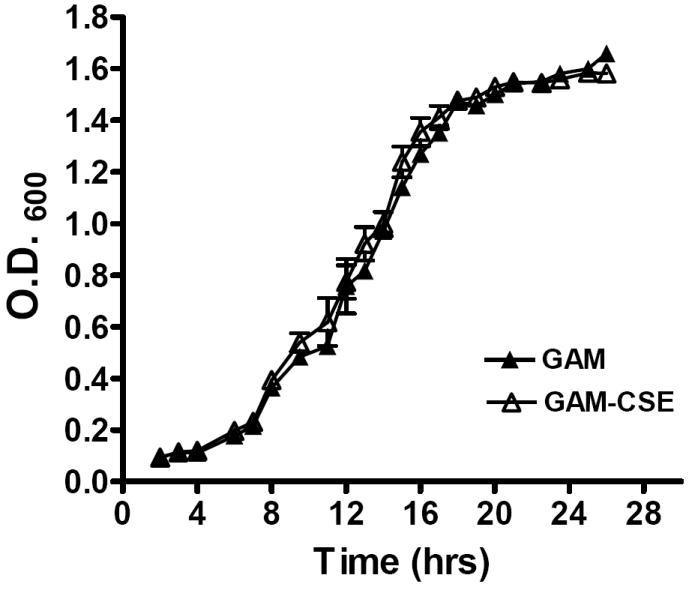
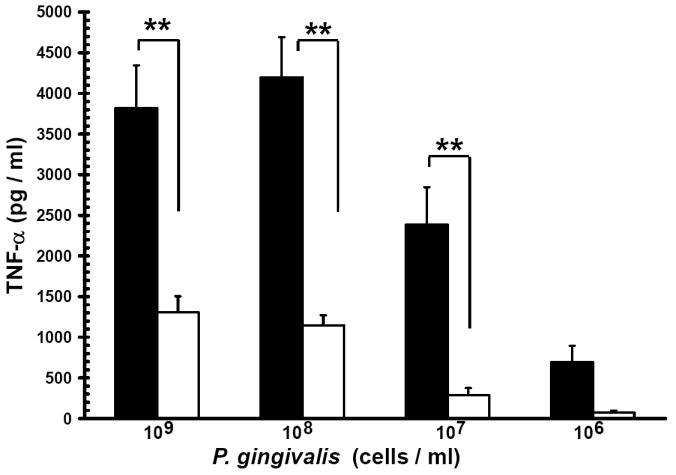
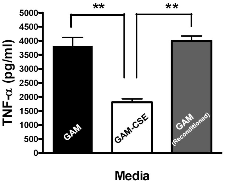
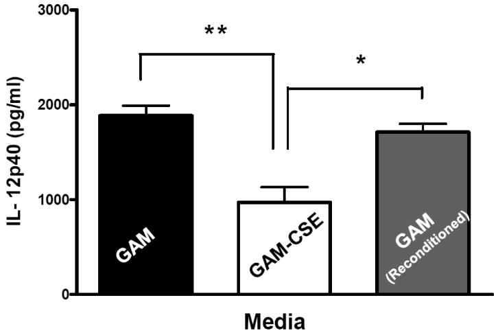
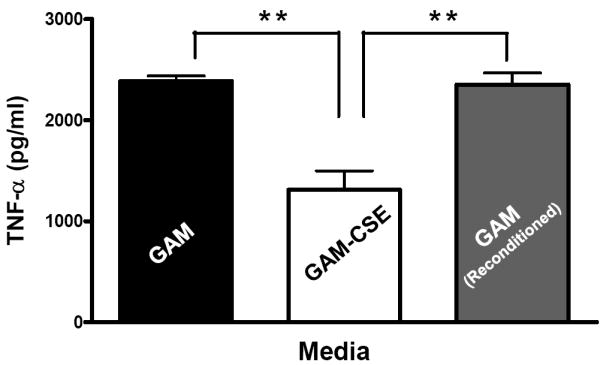
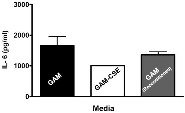
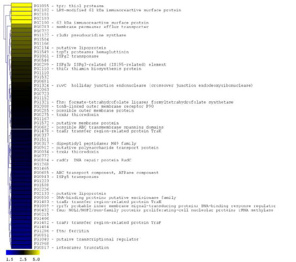
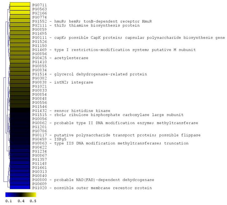
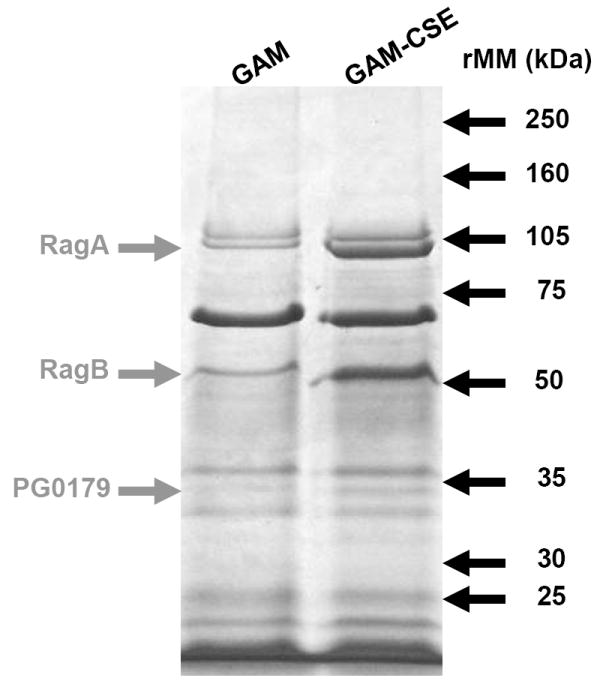
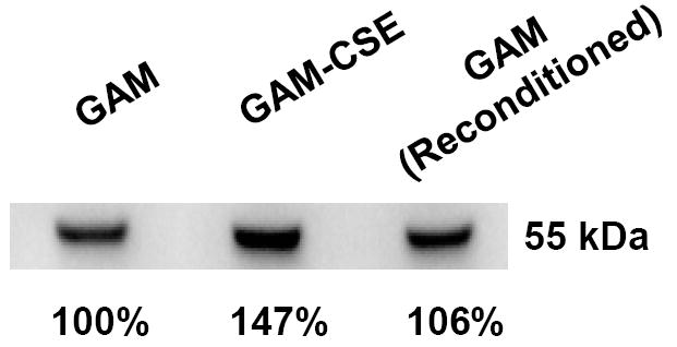
Smokers are more susceptible than non-smokers to persistent infection by Porphyromonas gingivalis, a causative agent of periodontitis. Patients who smoke exhibit increased susceptibility to periodontitis and are more likely to display severe disease and be refractory to treatment. Paradoxically, smokers demonstrate reduced clinical inflammation. We show that P. gingivalis cells exposed to cigarette smoke extract (CSE) induce a lower proinflammatory response (tumour necrosis factor-alpha, interleukin-6, interleukin-12 p40) from monocytes and peripheral blood mononuclear cells than do unexposed bacteria. This effect is reversed when CSE-exposed bacteria are subcultured in fresh medium without CSE. Using microarrays representative of the P. gingivalis genome, CSE-exposure resulted in differential regulation of 6.8% of P. gingivalis genes, including detoxification and oxidative stress-related genes; DNA repair genes; and multiple genes related to P. gingivalis virulence, including genes in the major fimbrial and capsular operons. Exposure to CSE also altered the expression of outer membrane proteins, most notably by inducing the virulence factors RagA and RagB, and a putative lipoprotein cotranscribed with the minor fimbrial antigen. Therefore, CSE represents an environmental stress to which P. gingivalis adapts by altering gene expression and outer membrane proteins. These changes may explain, in part, the altered virulence and host-pathogen interactions that have been documented in vivo in smokers with periodontal disease.


Figures

Figure 1
Figure 1. Typical growth curves of P. gingivalis W83 in CSE-conditioned and unconditioned medium
Growth of P. gingivalis was compared in GAM medium and GAM conditioned with CSE (500 ng ml-1 nicotine equivalents). Closed triangles represent growth in GAM. Open triangles represent growth in GAM-CSE. Error bars represent the mean (s.d.) of 3 experiments. There were no significant differences in the growth characteristics of P. gingivalis cultured in GAM or GAM-CSE (p > 0.05).
Figure 2
Figure 2. Reversible suppression of TNF-α release by human monocytes stimulated with CSE-treated P. gingivalis
(a) Reduced TNF-α release by human monocytes stimulated with CSE-treated P. gingivalis. Primary human monocytes (0.5 × 10⁶) were stimulated with 106 to 109 cells of control P. gingivalis (black bars) or GAM-CSE (grey bars) grown P. gingivalis cells (20h). Cell-free supernatants were harvested by centrifugation and levels of TNF-α were determined by ELISA. Error bars represent the mean (s.d.) of 3 experiments. ** Indicates statistical significance at p < 0.01. (b) Reconditioning of P. gingivalis in GAM rescues pro-inflammatory potential. Primary human monocytes (0.5 × 10⁶) were stimulated for 20 h with GAM cultured P. gingivalis; CSE-treated P. gingivalis; or P. gingivalis cells that were first grown in GAM-CSE for two passages then reconditioned in untreated GAM for 2 passages (107 P. gingivalis cells). Monocytes stimulated with reconditioned P. gingivalis produced levels of TNF-α that were similar to monocytes that were stimulated with bacteria from control cultures (not exposed to CSE). Error bars represent the mean (s.d.) of 3 experiments. ** Indicates statistical significance at p < 0.01.
Figure 3
Figure 3. Reversible suppression of multiple pro-inflammatory cytokines in human PBMCs stimulated with CSE-treated P. gingivalis
Primary human PBMCs (0.5 × 10⁶) were stimulated for 20 h with GAM cultured P. gingivalis; CSE-treated P. gingivalis; or P. gingivalis cells that were first grown in GAM-CSE for two passages then reconditioned in untreaαted GAM for 2 passages (107 P. gingivalis cells). PBMCs stimulated with reconditioned P. gingivalis produced levels of TNF-α that were similar to PBMCs that were stimulated with bacteria from control cultures (not exposed to CSE). Error bars represent the mean (s.d.) of 3 experiments. * Indicates statistical significance at p < 0.05. ** Indicates statistical significance at p < 0.01.
Figure 4
Figure 4. P. gingivalis genes differentially expressed following CSE exposure
The microarray data of Log2 ratios were uploaded onto MultipleExperiment viewer 4.1v (January 8, 2008 release) software and evaluated by t-test with p value based on ‘t’ distribution with significance (alpha cut-off) set at p <0.01. The subset of significant genes were divided into two groups based on i) upregulated genes (>1.5 fold increase) and ii) down regulated genes (<0.5 fold). The distance was calculated based on Euclidean distance with average linkage and the tree was generated based on hierarchical clustering. CSE-induced genes are shown in Figure 4a (significantly induced genes >1.5 fold compared to baseline; p < 0.01; genes are represented in yellow to blue colour with the lower limit = 1.5; midpoint value = 2.5; and upper limit = 5). CSE-suppressed genes are shown in Figure 4b (significantly down-regulated genes < 0.5 fold compared to baseline; p < 0.01; genes are represented in yellow to blue colour with the lower limit = 0.1; midpoint value = 0.4; and upper limit = 0.5).
Figure 5
Figure 5. Up-regulation of specific outer membrane protein (OMP) in CSE-exposed P. gingivalis
Outer membrane proteins of P. gingivalis grown in GAM and GAM-CSE were isolated by differential extraction with Triton X-100. 20μg total OMP extracts were run through a 4-15% gradient gel. MALDI-MS of excised bands followed by comparison with the ORF database of P. gingivalis W83 from The J. Craig Venter Institute identified the major CSE up-regulated bands, as shown.
Figure 6
Figure 6. CSE-induced RagB upregulation in P. gingivalis is reversible
A typical western blot of RagB in lysates of cells sequentially passaged in GAM, GAM-CSE and GAM, respectively, is shown. Culture in GAM-CSE resulted in an increase in relative RagB expression levels (147%). Relative RagB expression returned to near control levels on reconditioning in GAM.

References

    1. Aduse-Opoku J, Slaney JM, Hashim A, Gallagher A, Gallagher RP, Rangarajan M, et al. Identification and characterization of the capsular polysaccharide (K-antigen) locus of Porphyromonas gingivalis. Infect Immun. 2006;74:449–460. - PMC - PubMed
    1. Araco A, Gravante G, Sorge R, Araco F, Delogu D, Cervelli V. Wound infections in aesthetic abdominoplasties: the role of smoking. Plast Reconstr Surg. 2008;121:305e–310e. - PubMed
    1. Arcavi L, Benowitz NL. Cigarette smoking and infection. Arch Intern Med. 2004;164:2206–2216. - PubMed
    1. Barbour SE, Nakashima K, Zhang JB, Tangada S, Hahn CL, Schenkein HA, Tew JG. Tobacco and smoking: environmental factors that modify the host response (immune system) and have an impact on periodontal health. Crit Rev Oral Biol Med. 1997;8:437–460. - PubMed
    1. Bernard D. In: Molecular Mechanisms of Tobacco-Induced Diseases. Wang XL, Scott DA, editors. Nova Science; 2005.

Publication types

MeSH terms