Review
. 2009 Apr;30(12):2164-74.
doi: 10.1016/j.biomaterials.2008.12.084. Epub 2009 Jan 26.

Organ printing: tissue spheroids as building blocks

Affiliations
Review

Organ printing: tissue spheroids as building blocks

Vladimir Mironov et al. Biomaterials. 2009 Apr.

Abstract

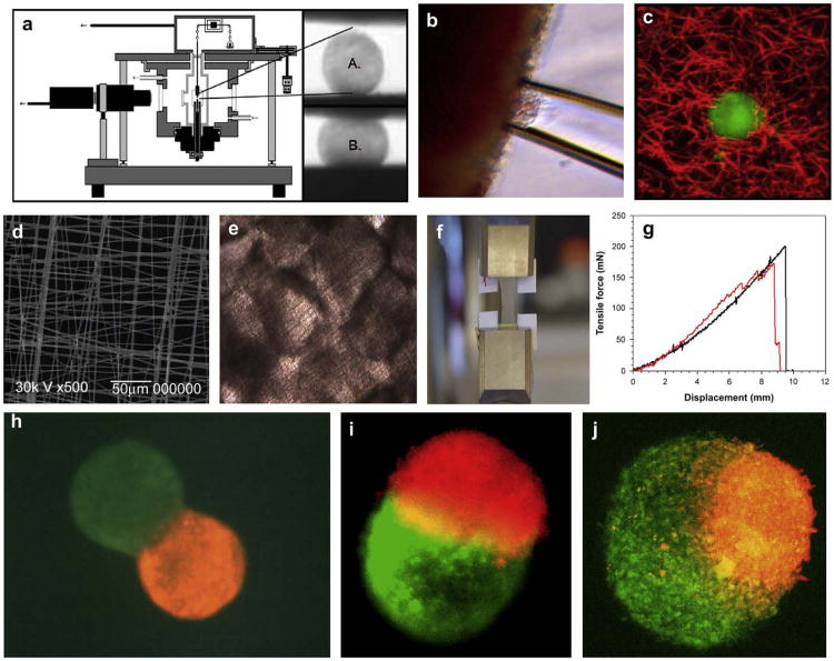
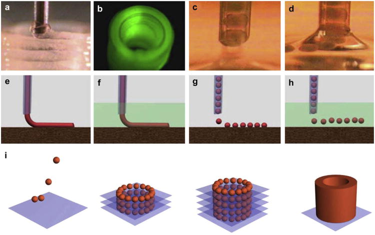
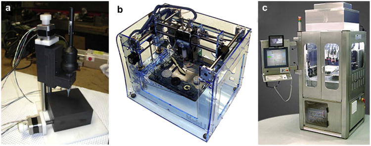
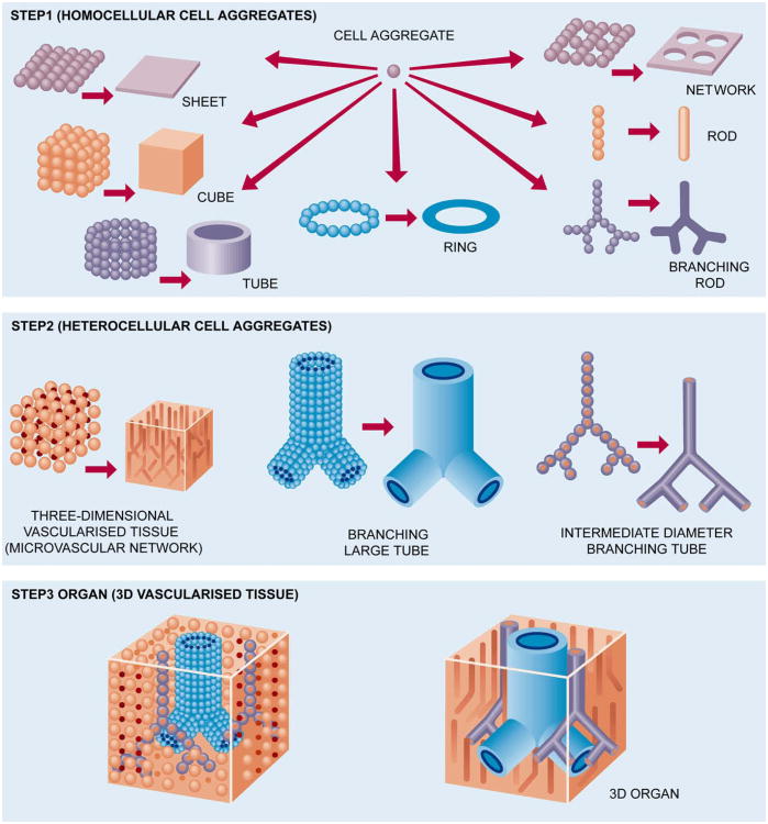
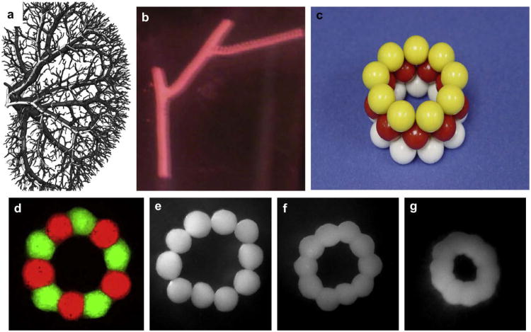
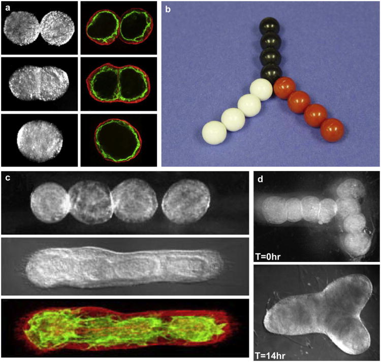
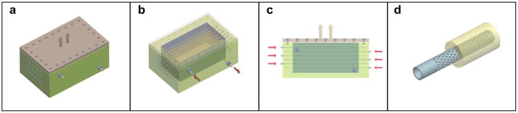
Organ printing can be defined as layer-by-layer additive robotic biofabrication of three-dimensional functional living macrotissues and organ constructs using tissue spheroids as building blocks. The microtissues and tissue spheroids are living materials with certain measurable, evolving and potentially controllable composition, material and biological properties. Closely placed tissue spheroids undergo tissue fusion - a process that represents a fundamental biological and biophysical principle of developmental biology-inspired directed tissue self-assembly. It is possible to engineer small segments of an intraorgan branched vascular tree by using solid and lumenized vascular tissue spheroids. Organ printing could dramatically enhance and transform the field of tissue engineering by enabling large-scale industrial robotic biofabrication of living human organ constructs with "built-in" perfusable intraorgan branched vascular tree. Thus, organ printing is a new emerging enabling technology paradigm which represents a developmental biology-inspired alternative to classic biodegradable solid scaffold-based approaches in tissue engineering.


Figures

Fig.1
Fig.1
Methods of biomechanical testing of tissue spheroids: a) tensiometry: experimental device and changing shape of tissue spheroid before (A) and after (B) tissue compression; b) aspiration assay; c) fluorescent microbead assay; d) SEM of elastic scaffold for tensile testing; e) fused tissue spheroids attached to elastic scaffold; f) tensile testing using tissue construct fabricated from fused tissue spheroids; g) example of force–displacement relationship for tissue construct fabricated from fused tissue spheroids; h) envelopment assay – initial step of tissue spheroid fusion; i) fusion of tissue spheroid with equal level of cohesion; j) fusion of tissue spheroid with different levels of tissue cohesion – lower cohesive tissue spheroid (green) is enveloping more cohesive tissue spheroid (red).
Fig. 2
Fig. 2
Principles of bioprinting technology: a) bioprinter (general view); b) multiple bioprinter nozzles; c) tissue spheroids before dispensing; d) tissue spheroids during dispensing; e) continuous dispensing in air; f) continuous dispensing in fluid; g) digital dispensing in air; h) digital dispensing in fluid; i) scheme of bioassembly of tubular tissue construct using bioprinting of self-assembled tissue spheroids illustrating sequential steps of layer-by-layer tissue spheroid deposition and tissue fusion process.
Fig. 3
Fig. 3
Bioprinters: a) 3D dispensing Laboratory Bioprinter – ‘LBP’ (designed by Neatco, Toronto, Canada in cooperation with MUSC Bioprinting Research Center, Charleston, SC); b) 3D robotic printer – ‘Fabber’ (designed by Cornell University, USA); c) 3D robotic industrial bioprinter – ‘BioAssembly Tool’ (designed by Sciperio/nScript, Orlando, USA).
Fig. 4
Fig. 4
Roadmap for organ printing.
Fig. 5
Fig. 5
Bioprinting of segments of intraorgan branched vascular tree using solid vascular tissue spheroids: a) kidney intraorgan vascular tree; b) bioprinted segment of vascular tree; c) physical model of bioassembly of tube-like vascular tissue construct using solid tissue spheroids; d) bioassembled ring-like vascular tissue constructs of tissue spheroids fabricated from human smooth muscle cells. Tissue spheroids are labeled with green and red fluorescent stains in order to demonstrate absence of cell mixing during tissue fusion process; e–g) sequential steps of morphological evolution of ring-like vascular tissue construct during tissue fusion process.
Fig. 6
Fig. 6
Bioprinting of segments of intraorgan branched vascular tree using uni-lumenal vascular tissue spheroids: a) fusion of uni-lumenal vascular tissue spheroids in hanging drop; b) physical model of fabrication of branched vascular segment from uni-lumenal vascular tissue spheroids; c) sequential steps of tissue fusion of vascular tissue spheroids placed in collagen type 1 hydrogel; d) fabrication branched vascular segments from uni-lumenal vascular tissue spheroids in collagen type 1 hydrogel (before and after tissue fusion process).
Fig. 7
Fig. 7
Computer simulation (using ‘Surface Evolver’ software) of morphology evolution of a tube-like vascular tissue construct during tissue fusion. The initial tube-like vascular tissue construct (a) is becoming shorter and more narrow (b). The hexagonal pattern of tissue spheroid packing is clearly demonstrated (b).
Fig. 8
Fig. 8
Scheme of the design of irrigation dripping tripled perfusion bioreactor: a) general view; b) view with opened lid; c) sectional view; d) design of removable microporous non-biodegradable tube.

References

    1. Griffith LG, Naughton G. Tissue engineering – current challenges and expanding opportunities. Science. 2002;295:1009–14. - PubMed
    1. Langer R, Vacanti JP. Tissue engineering. Science. 1993;260:920–6. - PubMed
    1. Mironov V, Boland T, Trusk T, Forgacs G, Markwald RR. Organ printing: computer-aided jet-based 3D tissue engineering. Trends Biotechnol. 2003;21:157–61. - PubMed
    1. Mironov V, Kasyanov V, Drake C, Markwald RR. Organ printing: promises and challenges. Regen Med. 2008;3:93–103. - PubMed
    1. Williams DF. On the mechanisms of biocompatibility. Biomaterials. 2008;29:2941–53. - PubMed

Publication types