. 2009 Feb 15;69(4):1268-72.
doi: 10.1158/0008-5472.CAN-08-3116. Epub 2009 Jan 27.

In vivo molecular imaging of cancer with a quenching near-infrared fluorescent probe using conjugates of monoclonal antibodies and indocyanine green

Affiliations

In vivo molecular imaging of cancer with a quenching near-infrared fluorescent probe using conjugates of monoclonal antibodies and indocyanine green

Mikako Ogawa et al. Cancer Res. .

Abstract

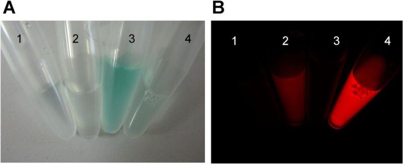
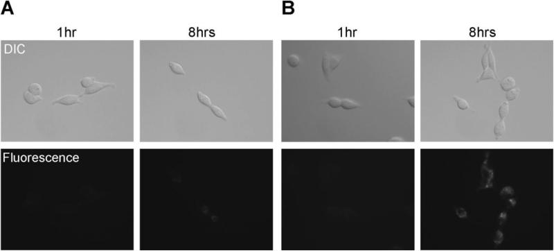
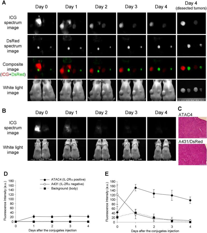
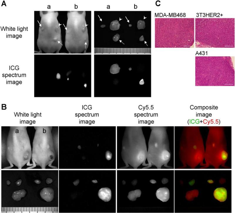
Near-infrared (NIR) fluorophores have several advantages over visible fluorophores, including improved tissue penetration and lower autofluorescence; however, only indocyanine green (ICG) is clinically approved. Its use in molecular imaging probes is limited because it loses its fluorescence after protein binding. This property can be harnessed to create an activatable NIR probe. After cell binding and internalization, ICG dissociates from the targeting antibody, thus activating fluorescence. ICG was conjugated to the antibodies daclizumab (Dac), trastuzumab (Tra), or panitumumab (Pan). The conjugates had almost no fluorescence in PBS but became fluorescent after SDS and 2-mercaptoethanol, with a quenching capacity of 10-fold for 1:1 conjugates and 40- to 50-fold for 1:5 conjugates. In vitro microscopy showed activation within the endolysosomes in target cells. In vivo imaging in mice showed that CD25-expressing tumors were specifically visualized with Dac-ICG. Furthermore, tumors overexpressing HER1 and HER2 were successfully characterized in vivo by using Pan-ICG(1:5) and Tra-ICG(1:5), respectively. Thus, we have developed an activatable NIR optical probe that "switches on" only in target cells. Because both the antibody and the fluorophore are Food and Drug Administration approved, the likelihood of clinical translation is improved.


Figures

Figure 1
Figure 1
The bright field (A) and fluorescence (B) images of antibody-ICG conjugates. 1, 2: Zen-ICG(1:1), 3, 4: Zen-ICG(1:1). 1, 3: in PBS, 2, 4: SDS and 2-ME added condition. In PBS, all the conjugates have no fluorescence. The fluorescence was activated by SDS and 2-ME treatment.
Figure 2
Figure 2
Fluorescence microscopy. 3T3/HER2+ cells were incubated with Tra-ICG(1:1) (A) or Tra-ICG(1:5) (B) for 1hr or 8hr. The fluorescent signal on the cell surface was not observed after 1hr incubation. The fluorescent signal was detected after internalization into the cells by 8hr incubation. The signal was higher for Tra-ICG(1:5) than Tra-ICG(1:1).
Figure 3
Figure 3
In vivo and ex vivo spectral fluorescence images with ATAC4 (left dorsum) and A431/DsRed (right dorsum) tumor bearing mice (A, B). A: dorsal images, B: ventral images. Dac-ICG(1:5) injected mouse is on the left side and Dac-ICG(1:1) injected mouse is on the right side. The target tumor (ATAC4) was visualized by both conjugates, but the brightness was higher for Dac-ICG(1:5). The non-target tumor (A431/DsRed) was not detected by either conjugate. C: H&E staining of dissected tumors (bar = 200 μm). Histologic findings of both tumors were identical. High initial liver uptake was observed for Dac-ICG(1:5), but the clearance was rapid. The time course of ICG fluorescence intensities after intravenous injection of Dac-ICG(1:1) (D) or Dac-ICG(1:5) (E). The fluorescence intensity was increased only in the target tumor (ATAC4).
Figure 4
Figure 4
A: Fluorescence imaging with HER1 positive and HER2 positive tumor bearing mice. Arrow head; 3T3/HER2 tumors (HER2 positive), solid arrow; MDA-MB468 tumors (HER1 positive), dashed arrow; A431 (HER1 positive) tumors. a; Tra-ICG(1:5) injected mouse, b; Pan-ICG(1:5) injected mouse. The images were obtained 4 days after the injection. Only the target specific tumor was detected and tumor characterization was successful. B: Human polyclonal human IgG-Cy5.5 was co-injected with Tra-ICG(1:5) (a) or Pan-ICG(1:5) (b) to validate specific accumulation. Only the target tumor was visualized with each specific ICG conjugated antibody, however, all tumors were detected with IgG-Cy5.5. C: H&E staining of dissected tumors (bar = 200 μm).

References

    1. Sharma R, Wendt JA, Rasmussen JC, Adams KE, Marshall MV, Sevick-Muraca EM. New horizons for imaging lymphatic function. Ann N Y Acad Sci. 2008;1131:13–36. - PMC - PubMed
    1. Weissleder R. A clearer vision for in vivo imaging. Nat Biotechnol. 2001;19:316–7. - PubMed
    1. Sakka SG. Assessing liver function. Curr Opin Crit Care. 2007;13:207–14. - PubMed
    1. Dzurinko VL, Gurwood AS, Price JR. Intravenous and indocyanine green angiography. Optometry. 2004;75:743–55. - PubMed
    1. Urano Y, Asanuma D, Hama Y, et al. Selective molecular imaging of viable cancer cells with pH-activatable fluorescence probes. Nat Med. 2008 in press. - PMC - PubMed

Publication types