. 2009 Feb 1;69(3):1156-65.
doi: 10.1158/0008-5472.CAN-08-2917. Epub 2009 Jan 27.

Suppression of cFLIP by lupeol, a dietary triterpene, is sufficient to overcome resistance to TRAIL-mediated apoptosis in chemoresistant human pancreatic cancer cells

Affiliations

Suppression of cFLIP by lupeol, a dietary triterpene, is sufficient to overcome resistance to TRAIL-mediated apoptosis in chemoresistant human pancreatic cancer cells

Imtiyaz Murtaza et al. Cancer Res. .

Abstract

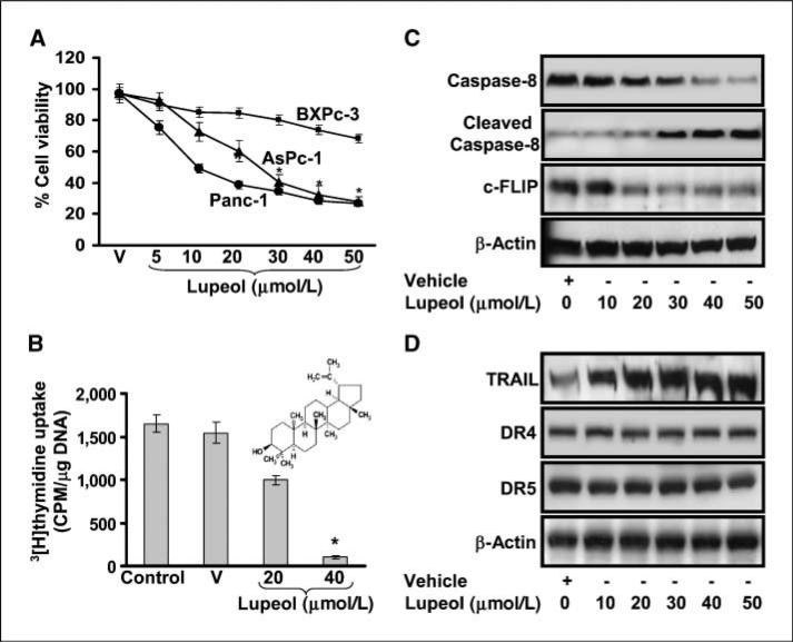
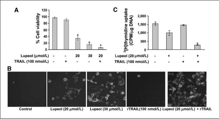
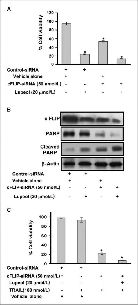
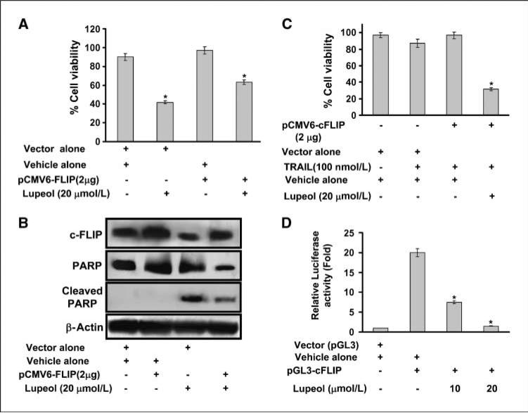
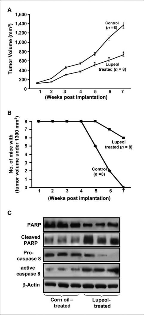
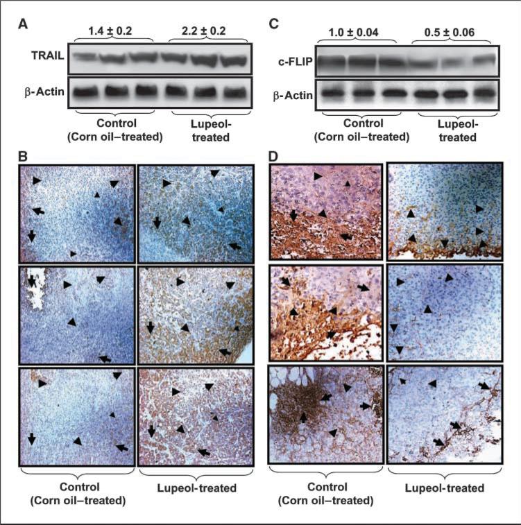
Overexpression of cellular FLICE-like inhibitory protein (cFLIP) is reported to confer chemoresistance in pancreatic cancer (PaC) cells. This study was designed to investigate the effect of lupeol, a dietary triterpene, on (a) apoptosis of tumor necrosis factor-related apoptosis-inducing ligand (TRAIL) therapy-resistant PaC cells overexpressing cFLIP and (b) growth of human pancreatic tumor xenografts in vivo. The effect of lupeol treatment on proliferation and TRAIL/caspase-8/cFLIP machinery in PaC cells was investigated. Next, cFLIP-overexpressing and cFLIP-suppressed cells were tested for sensitivity to recombinant TRAIL therapy in the presence of lupeol. Further, athymic nude mice implanted with AsPC-1 cells were treated with lupeol (40 mg/kg) thrice a week and surrogate biomarkers were evaluated in tumors. Lupeol alone treatment of cells caused (a) decrease in proliferation, (b) induction of caspase-8 and poly(ADP-ribose) polymerase cleavage, and (c) down-regulation of transcriptional activation and expression of cFLIP. Lupeol was observed to increase the TRAIL protein level in cells. Lupeol significantly decreased the viability of AsPC-1 cells both in cFLIP-suppressed cells and in cFLIP-overexpressing cells. Lupeol significantly sensitized chemoresistant PaC cells to undergo apoptosis by recombinant TRAIL. Finally, lupeol significantly reduced the growth of human PaC tumors propagated in athymic nude mice and caused modulation of cFLIP and TRAIL protein levels in tumors. Our findings showed the anticancer efficacy of lupeol with mechanistic rationale against highly chemoresistant human PaC cells. We suggest that lupeol, alone or as an adjuvant to current therapies, could be useful for the management of human PaC.


Figures

Figure 1
Figure 1
Effect of lupeol on cell viability, proliferation, activation of caspase-8, and the expression level of cFLIP, TRAIL, and death receptors in AsPC-1cells. A, viability of human PaC AsPC-1, BxPC-3, and PANC-1 cells treated with lupeol (5–50 μmol/L for 48 h) as measured by MTT assay. Each concentration of lupeol was repeated in 10 wells. The values are represented as percent viable cells, with vehicle (alcohol + DMSO)-treated cells regarded as 100% viable. Points, mean of three independent experiments; bars, SE. B, histogram showing the rate of [3H]thymidine uptake in AsPC-1 cells treated with lupeol. Cells were subjected to lupeol treatment (20–40 μmol/L) for 48 h, the last 16 h of which was in the presence of [3H]thymidine (0.5 μCi/mL). Columns, mean of three independent experiments; bars, SE. *, P < 0.05. Inset, structure of lupeol [Lup-20(29)-en-3β-ol]. C, effect of lupeol (0–50 μmol/L) treatment on the expression levels of procaspase-8 and cleaved caspase-8 and on the protein expression of cFLIP. D, effect of lupeol (0–50 μmol/L) treatment on the expression levels of TRAIL, DR4, and DR5 proteins as determined by immunoblot analysis. Representative of three independent experiments with similar results. The details are described in Materials and Methods. V, vehicle (alcohol + DMSO).
Figure 2
Figure 2
Effect of lupeol and rTRAIL treatment, alone and in combination (Lupeol + rTRAIL), on cell viability, proliferation, and apoptosis of AsPC-1cells. A, histogram representing the viability of cells treated with specified concentrations of lupeol (20–30 μmol/L) and rTRAIL (100 nmol/L). Cells were treated with lupeol for 24 h and were then exposed to rTRAIL for 6 h. After TRAIL treatment, cell viability was assessed by MTT assay. Each concentration of lupeol was repeated in 10 wells. The values are represented as percent viable cells, with vehicle (alcohol + DMSO)–treated cells regarded as 100% viable. Columns, mean of three independent experiments; bars, SE. B, representative photomicrographs showing induction of apoptosis in AsPC-1 cells treated with lupeol and rTRAIL alone and in combination. Green and red fluorescence indicate Annexin V and propidium iodide staining. Representative of three independent experiments with similar results. C, histogram showing the rate of [3H]thymidine uptake in AsPC-1cells treated with lupeol and rTRAIL alone and in combination. Cells were subjected to lupeol treatment (20 μmol/L) for 48 h, the last 16 h of which was in the presence of [3H]thymidine (0.5 μCi/mL). Columns, mean of three independent experiments; bars, SE. *, P < 0.01. The details are described in Materials and Methods.
Figure 3
Figure 3
Effect of lupeol treatment on cell viability and on the expression of cFLIP and PARP cleavage in cFLIP-silenced AsPC-1cells. A, histogram representing the effect of lupeol on the growth of cFLIP-silenced AsPC-1cells as determined by MTT assay. Cells were transfected with cFLIP siRNA or scrambled siRNA (control) by electroporation. Twelve hours posttransfection, cells were treated with specified concentrations of lupeol and cell viability was determined. The values represent percent viable cells, with vehicle-treated cells regarded as 100% viable. Columns, mean percent viable cells of three independent experiments; bars, SE. The details are described in Materials and Methods. B, representative immunoblots of cFLIP and PARP protein cleavage. Cells were transfected with cFLIP siRNA or scrambled siRNA (control) by electroporation. At 12 h posttransfection, cells were treated with specified concentrations of lupeol. After 48 h, cells were harvested and cell lysates analyzed for cFLIP and PARP cleavage. Equal loading was confirmed by stripping immunoblots and reprobing them for β-actin. C, histogram representing the effect of rTRAIL and lupeol alone and in combination on the growth of cFLIP-silenced AsPC-1cells. Cells were transfected with cFLIP siRNA or scrambled siRNA (control) by electroporation. Twelve hours posttransfection, cells were treated with specified concentrations of rTRAIL, lupeol, or their combination. After 48 h, cell viability was determined by MTT assay. The values represent percent viable cells, with vehicle-treated cells regarded as 100% viable. Columns, mean percent viable cells of three independent experiments; bars, SE. The details are described in Materials and Methods.
Figure 4
Figure 4
Effect of lupeol treatment on cell viability and on the expression of cFLIP and PARP cleavage in AsPC-1cells transfected with cFLIP-overexpressing plasmid. A, histogram representing the effect of lupeol on the growth of cFLIP-overexpressing AsPC-1cells. Cells were transfected with pCMV6-cFLIP or pCMV6 vector alone (control) by electroporation. Twelve hours posttransfection, cells were treated with specified concentrations of lupeol and cell viability was determined by MTT assay. The values represent percent viable cells, with vehicle-treated cells regarded as 100% viable. Columns, mean percent viable cells of three independent experiments; bars, SE. The details are described in Materials and Methods. B, representative immunoblots of cFLIP and PARP protein cleavage in cFLIP-overexpressing AsPC-1cells. Cells were transfected with pCMV6-cFLIP or pCMV6 vector alone (control) by electroporation. Twelve hours posttransfection, cells were treated with specified concentrations of lupeol; cell lysates were prepared and analyzed for cFLIP and PARP cleavage by Western blot analysis. Equal loading was confirmed by stripping immunoblots and reprobing them for β-actin. Representative immunoblots of three independent experiments with similar results. C, histogram representing the effect of rTRAIL and lupeol alone and in combination on the growth of cFLIP-overexpressing AsPC-1cells. Cells were transfected with pCMV6-cFLIP or pCMV6 vector alone (control) by electroporation. Twelve hours posttransfection, cells were treated with specified concentrations of rTRAIL and lupeol alone and in combination and cell viability was determined by MTT assay. The values represent percent viable cells, with vehicle-treated cells regarded as 100% viable. Columns, mean percent viable cells of three independent experiments; bars, SE. The details are described in Materials and Methods. D, histogram representing the effect of lupeol treatment on the transcriptional activation of cFLIP in AsPC-1cells. Cells were transfected with pGL3-cFLIP-luc for 24 h. Renilla luciferase was used as an internal control. In addition, controls were transfected with the same amount of empty vectors. Twelve hours posttransfection, fresh medium was added with lupeol and incubated for 24 h. The cells were then harvested and transcriptional activity was measured in quadruplicates as described in Materials and Methods.
Figure 5
Figure 5
Effect of lupeol administration on the tumorigenicity of AsPC-1cells and the expression level of known apoptotic markers PARP and caspase-8 under in vivo conditions. A, graphical representation of data showing the effect of lupeol treatment on the growth of tumors from AsPC-1cells implanted in athymic nude mice. The growth was measured in terms of average volume of tumors as a function of time. Points, mean; bars, SD. *, P < 0.05, versus the control group. B, graphical representation of the data depicting the number of mice remaining with tumor volumes <1,300 mm3 after treatment with corn oil alone or lupeol for the indicated weeks. C, effect of lupeol administration on the expression levels of PARP cleavage, procaspase-8, and active caspase-8 in AsPC-1cell–derived tumors excised at the 49th day posttreatment as determined by immunoblot analysis. Equal loading was confirmed by stripping the membrane and reprobing them for β-actin. Representative immunoblot of three samples from each group. The details are described in Materials and Methods.
Figure 6
Figure 6
Effect of lupeol administration on the expression level of cFLIP and TRAIL proteins in AsPC-1cell–derived tumors in athymic nude mice. A, effect of lupeol treatment on the expression level of TRAIL in AsPC-1cell–derived tumors excised at the 49th day posttreatment as determined by immunoblot analysis. Equal loading was confirmed by stripping the membrane and reprobing them for β-actin. Representative immunoblot of three samples from each group. Values above the immunoblots represent mean relative densities of the bands normalized to β-actin ± SE. B, representative photomicrographs (magnification, × 200) showing immunohistochemical staining for TRAIL in tumor sections of corn oil–treated and lupeol-treated mice. Arrows, regions exhibiting immunoreactivity for TRAIL protein. The immunostaining data were confirmed in two or more specimens of each group. C, effect of lupeol treatment on the expression levels of cFLIP in AsPC-1cell–derived tumors excised at the 49th day posttreatment as determined by immunoblot analysis. Equal loading was confirmed by stripping the membrane and reprobing them for β-actin. Representative immunoblot of three samples from each group. Values above the immunoblots represent mean relative densities of the bands normalized to β-actin; bars, SE. D, representative photomicrographs (magnification, × 200) showing immunohistochemical staining for cFLIP in tumor sections of corn oil–treated and lupeol-treated mice. Arrows, regions exhibiting immunoreactivity for cFLIP protein. The immunostaining data were confirmed in three specimens from each group. The details are described in Materials and Methods.

Similar articles

Cited by

References

    1. Jemal A, Siegel R, Ward E, et al. Cancer statistics, 2008. CA Cancer J Clin. 2008;58:71–96. - PubMed
    1. Garcea G, Dennison AR, Pattenden CJ, Neal CP, Sutton CD, Berry DP. Survival following curative resection for pancreatic ductal adenocarcinoma. A systematic review of the literature. JOP. 2008;9:99–132. - PubMed
    1. Mimeault M, Brand RE, Sasson AA, Batra SK. Recent advances on the molecular mechanisms involved in pancreatic cancer progression and therapies. Pancreas. 2005;31:301–16. - PubMed
    1. Westphal S, Kalthoff H. Apoptosis: targets in pancreatic cancer. Mol Cancer. 2003;2:6. - PMC - PubMed
    1. Xiong HQ. Molecular targeting therapy for pancreatic cancer. Cancer Chemother Pharmacol. 2004;54:69–77. - PubMed

Publication types

MeSH terms