. 2009 Mar;136(5):833-41.
doi: 10.1242/dev.028845. Epub 2009 Jan 28.

Neurovascular development uses VEGF-A signaling to regulate blood vessel ingression into the neural tube

Affiliations

Neurovascular development uses VEGF-A signaling to regulate blood vessel ingression into the neural tube

Jennifer M James et al. Development. 2009 Mar.

Abstract

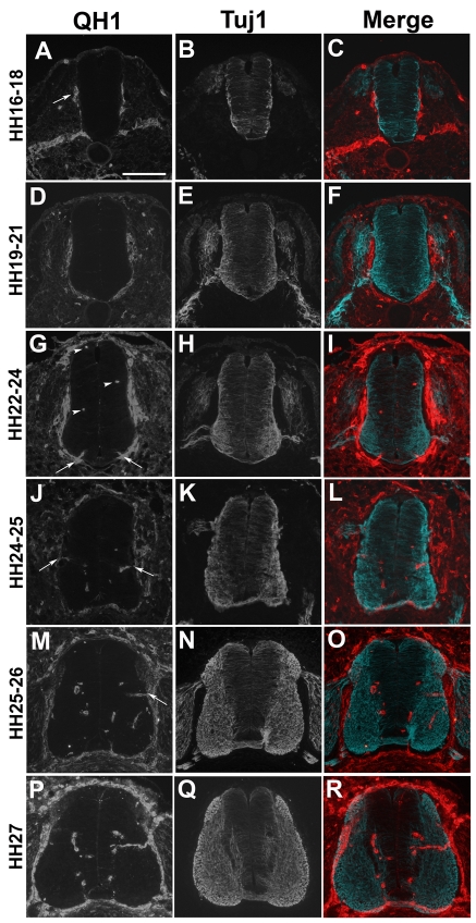
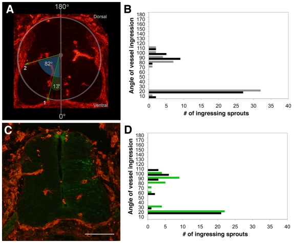
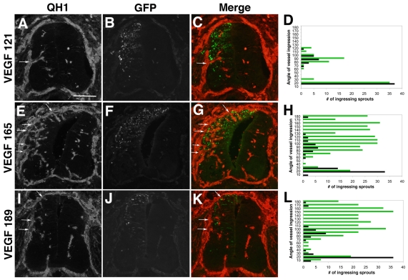
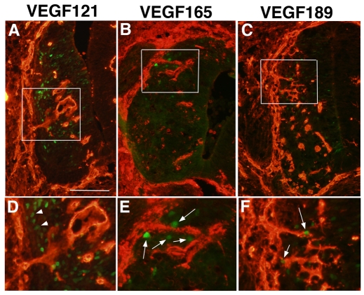
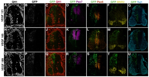
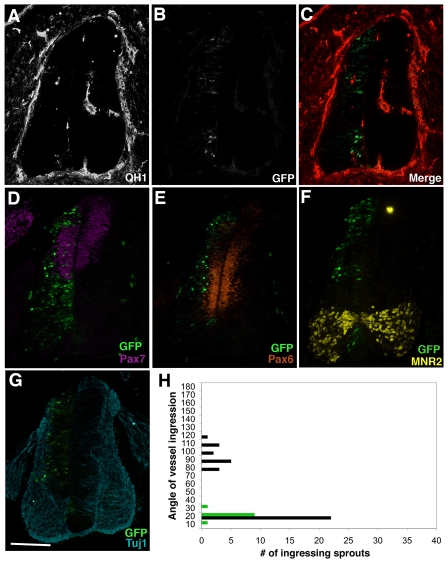
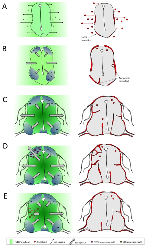
Neurovascular development requires communication between two developing organs, the neuroepithelium and embryonic blood vessels. We investigated the role of VEGF-A signaling in the embryonic crosstalk required for ingression of angiogenic vessel sprouts into the developing neural tube. As the neural tube develops, blood vessels enter at specific points medially and ventrally from the surrounding perineural vascular plexus. Localized ectopic expression of heparin-binding VEGF165 or VEGF189 from the developing avian neural tube resulted in supernumerary blood vessel ingression points and disrupted vessel patterning. By contrast, localized ectopic neural expression of non-heparin-binding VEGF121 did not produce supernumerary blood vessel ingression points, although the vessels that entered the neural tube became dysmorphogenic. Localized loss of endogenous VEGF-A signaling in the developing neural tube via ectopic expression of the VEGF inhibitor sFlt-1 locally blocked blood vessel ingression. The VEGF pathway manipulations were temporally controlled and did not dramatically affect neural tube maturation and dorsal-ventral patterning. Thus, neural-derived VEGF-A has a direct role in the spatially localized molecular crosstalk that is required for neurovascular development and vessel patterning in the developing neural tube.


Figures

Fig. 1.
Fig. 1.
Formation of the PNVP and vessel ingression coordinate with neural differentiation in the quail neural tube. HH stage 16-27 Japanese quail embryos were sectioned in the transverse plane at the thoracic level and stained with either QH1 (red, A,D,G,J,M,P) to label blood vessels or Tuj1 (β tubulin III, blue, B,E,H,K,N,Q) to label differentiated neurons. Merged images (C,F,I,L,O,R) represent two super-imposed, adjacent 12 μm sections at the upper limb level. (A-C) At stage HH 16-18, initiation of PNVP formation correlated with the start of neuronal differentiation and migration of Tuj1-positive neurons to the pial surface of the neural tube. (D-F) At stage HH 19-21, the PNVP continued to develop around the ventral neural tube, whereas motoneurons extended axonal projections from the motor horn, and DRG axons innervated the neural tube at the dorsal root entry zone. (G-I) By stage HH 22-24, PNVP formation was complete. Single QH1-positive angioblasts were noted dorsally and medially (arrowheads), and ventral angiogenic sprouts were seen (arrows) adjacent to the floor plate. (J-L) At stage HH 24-25, angiogenic sprouts from the PNVP formed mediolaterally (arrows) along the dorsal-ventral axis of the neural tube (this vessel ingression site was maintained at later stages, see M and P). (M-O) At stage HH 25-26, both ventral and medial (arrow) vessel ingression sites were noted, along with continued differentiation of Tuj1-positive neurons. (P-R) By stage HH 27, the amount of Tuj1-positive neurons increased, while the vessel ingression pattern established at earlier stages was maintained. Scale bar: 100 μm.
Fig. 2.
Fig. 2.
Quantitative analysis of angiogenic sprouting into the developing neural tube reveals stereotypical ingression points. Unperturbed quail embryos and embryos whose neural tubes were electroporated with eGFP control DNA at HH stage 16-18 were serially sectioned at stage HH 25-26 through the upper limb. Every sixth 12 μm section was stained for QH1 (red). Fourteen images were analyzed for each embryo as described in the Materials and methods. (A) Unperturbed quail embryo section stained with QH1 to illustrate blood vessel analysis strategy. (B) Total number of angiogenic sprouts within the left (gray) and right (black) neural tube halves of five unperturbed embryos. There were concentrations of ingression points between 0-20° (ventral ingression points) and 70-110° (medial ingression points). (C) Representative image of a quail neural tube electroporated with eGFP DNA (electroporated side to the right). (D) Total number of ingressing angiogenic sprouts within the neural tubes of five control embryos electroporated with eGFP DNA (green); untransfected control contralateral neural tube side (black). Scale bar: 100 μm.
Fig. 3.
Fig. 3.
Ectopic expression of heparin-binding VEGF isoforms induces supernumerary vessel ingression points into the developing neural tube. Quail neural tubes were electroporated with hVEGF121-GFP, hVEGF165-GFP or hVEGF189-GFP DNAs (green, panels B, F, J) on day 3 (HH 16-18) and harvested 48 hours later (HH 25-26). Transverse sections were stained with QH1 antibody (red, panels A,E,I) to visualize vessels, and five embryos from each group were analyzed as described (panels D,H,L; green lines, total ingression points for ectopic VEGF-expressing sides of neural tubes at each 10° of arc; black lines, total ingression points for contralateral control sides of the neural tubes at each 10° of arc). C, G and K are a merge of red (QH1) and green (eGFP) channels. (A-C) Quail neural tubes electroporated with hVEGF121 DNA displayed a grossly normal distribution of angiogenic ingression points along the dorsoventral axis of the ectopic VEGF-expressing side of the neural tube (arrows in A,C). (D) The quantitative analysis showed no change in the distribution of ingression points for sprouts between the control (black) and VEGF121-expressing (green) sides of the neural tube, and a slight increase in the frequency of ingression points in the medial region of the VEGF-expressing side of the neural tubes (n=5 embryos). (E-G) Quail neural tubes electroporated with hVEGF165 DNA had ectopic dorsal sprouts (arrows in E,G). (H) The quantitative analysis showed increased distribution and frequency of vessel ingression points in the dorsal region of the hVEGF165-expressing side of the neural tube (green), where ectopic expression is localized (n=5 embryos). (I-K) Quail neural tubes electroporated with hVEGF189 DNA had ectopic dorsal sprouts (arrows in I,K). (L) The quantitative analysis showed increased distribution and frequency of vessel ingression points in the dorsal region of the hVEGF189-expressing side of the neural tube, where ectopic expression is localized (n=5 embryos). Scale bar: 100 μm.
Fig. 4.
Fig. 4.
Localized ectopic expression of heparin-binding VEGF-A isoforms in the developing neural tube correlates with supernumerary vessel ingression points. Neural tubes processed for QH1 (red) and eGFP (green, reporter for ectopic VEGF-A isoform expression) were examined for the relationship between vessel ingression points and ectopic VEGF-A isoform expression. (A-C) Lower power views to show location of normal (A) or supernumerary (B,C) vessel sprout ingressions on the dorsoventral axis of the neural tube. (D-F) Higher magnification of the boxed areas in A-C. Several eGFP-positive cells that ectopically express heparin-binding hVEGF165 or hVEGF189 are close to the supernumerary vessel sprouts (arrows in E,F), whereas numerous eGFP-positive cells that ectopically express hVEGF121 (arrowheads in D) do not induce supernumerary vessel ingression points. Scale bar: 100 μm in A-C; 50 μm in D-F.
Fig. 5.
Fig. 5.
Neural patterning is not perturbed in neural tubes that ectopically express VEGF-A isoforms. (A-U) Quail neural tubes were electroporated with (A-G) hVEGF121, (H-N) hVEGF165 and (O-U) hVEGF189 on day 3 (HH 16-18), and harvested 48 hours later (HH 25-26). Neural tubes were sectioned and adjacent sections were stained with antibodies to: QH1 (red, A,H,O,C,J,Q) to visualize vessels; Pax7 (purple, D,K,R) to visualize dorsal neural precursors; Pax6 (orange, E,L,S) to visualize medial neural precursors; MNR2 (yellow, F,M,T) to visualize ventral motoneuron precursors; and Tuj1 (blue, G,N,U) to visualize differentiated neurons. (B,I,P) eGFP expression (green) illustrates the neural tube side expressing ectopic VEGF-A isoforms (left) versus the control contralateral side (right); (C-G,J-N,Q-U) merges of marker and eGFP channels for each section. Scale bar: 100 μm.
Fig. 6.
Fig. 6.
VEGF signaling from the neural tube is required for blood vessel ingression. Quail neural tubes were electroporated with sFlt1-GFP and analyzed as previously described. (A-C) No medial vessel ingression and little ventral vessel ingression was seen in areas of the neural tube that were eGFP positive. (D-G) Neural patterning is not detectably perturbed on the electroporated side of the neural tube (left) based on Pax7 (D, purple), Pax6 (E, orange), MNR2 (F, yellow) and Tuj1 (G, blue) expression patterns. (H) Quantitative analysis of five electroporated neural tubes showed no medial and few ventral vessel ingression points in areas of localized sFlt1 expression (green), compared with the control contralateral side (black) (n=5 embryos). Scale bar: 100 μm.
Fig. 7.
Fig. 7.
Model of blood vessel ingression into the developing neural tube. The model covers events of neurovascular patterning between stages HH 16 and HH 26 in the avian embryo, and also shows the VEGF-A perturbations analyzed in this study. (A) At early stages (HH 16-18), VEGF-A isoforms (121, 165, 189) expressed by the developing neural tube set up a gradient that leads to angioblast migration from the lateral plate and presomitic mesoderm to form the PNVP. At this stage, blood vessel ingression does not occur because of insufficient levels of neural tube-derived VEGF-A. (B) At later stages (HH 22-25), increased levels of VEGF165 and VEGF189 are required for blood vessel ingression, but negative patterning cues that are co-expressed prevent ingression except at specific medial and ventral points. (C) By stage HH 26 there are obvious stereotypical blood vessel ingression points medially and ventrally, whereas angioblasts migrate in dorsally. (D) Neural tubes electroporated with VEGF165 or VEGF 189 show ectopic ingression in normally avascular dorsal areas on the electroporated side. (E) Neural tubes electroporated with sFlt-1 do not have ingression at the normal medial site on the electroporated side. For each set of panels, the left side demonstrates the signals and the right side demonstrates the vessel patterning outcome. Symbols are described in the key below the figure.

Similar articles

Cited by

References

    1. Aitkenhead, M., Christ, B., Eichmann, A., Feucht, M., Wilson, D. J. and Wilting, J. (1998). Paracrine and autocrine regulation of vascular endothelial growth factor during tissue differentiation in the quail. Dev. Dyn. 212, 1-13. - PubMed
    1. Ambler, C. A., Nowicki, J. L., Burke, A. C. and Bautch, V. L. (2001). Assembly of trunk and limb blood vessels involves extensive migration and vasculogenesis of somite-derived angioblasts. Dev. Biol. 234, 352-364. - PubMed
    1. Ambler, C. A., Schmunk, G. A. and Bautch, V. L. (2003). Stem cell-derived endothelial cells/progenitors migrate and pattern in the embryo using the VEGF signaling pathway. Dev. Biol. 257, 205-219. - PubMed
    1. Carmeliet, P. and Tessier-Lavigne, M. (2005). Common mechanisms of nerve and blood vessel wiring. Nature 436, 193-200. - PubMed
    1. Carmeliet, P., Ferreira, V., Breier, G., Pollefeyt, S., Kieckens, L., Gertsenstein, M., Fahrig, M., Vandenhoeck, A., Harpal, K., Eberhardt, C. et al. (1996). Abnormal blood vessel development and lethality in embryos lacking a single VEGF allele. Nature 380, 435-439. - PubMed

Publication types

MeSH terms