. 2009 Mar;136(5):843-53.
doi: 10.1242/dev.027805. Epub 2009 Jan 28.

Coordinate integrin and c-Met signaling regulate Wnt gene expression during epithelial morphogenesis

Affiliations

Coordinate integrin and c-Met signaling regulate Wnt gene expression during epithelial morphogenesis

Yingjie Liu et al. Development. 2009 Mar.

Abstract

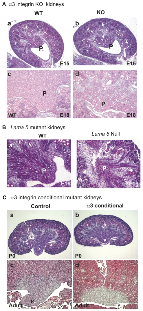
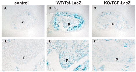
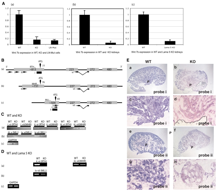
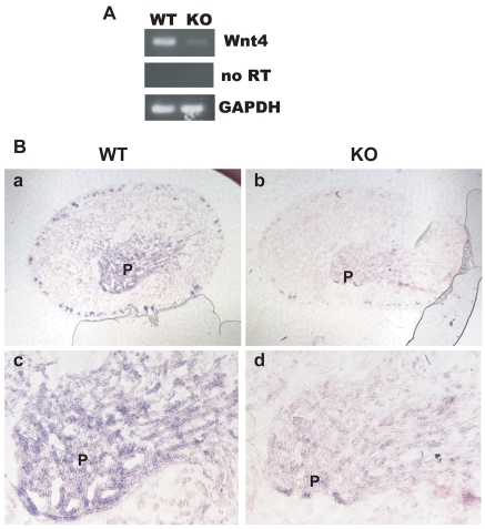
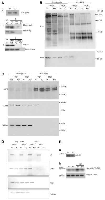
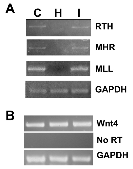
Integrin receptors for the extracellular matrix and receptor tyrosine kinase growth factor receptors represent two of the major families of receptors that transduce into cells information about the surrounding environment. Wnt proteins are a major family of signaling molecules that regulate morphogenetic events. There is presently little understanding of how the expression of Wnt genes themselves is regulated. In this study, we demonstrate that alpha3beta1 integrin, a major laminin receptor involved in the development of the kidney, and c-Met, the receptor for hepatocyte growth factor, signal coordinately to regulate the expression of Wnt7b in the mouse. Wnt signals in turn appear to regulate epithelial cell survival in the papilla of the developing kidney, allowing for the elongation of epithelial tubules to form a mature papilla. Together, these results demonstrate how signals from integrins and growth factor receptors can be integrated to regulate the expression of an important family of signaling molecules so as to regulate morphogenetic events.


Figures

Fig. 1.
Fig. 1.
Histology of α3β1 integrin-deficient or Lama5-deficient kidney papillae. (A) Histology and proliferation of (a,c) wild-type (WT) and (b,d) α3 integrin KO kidneys. (a,b) E15; (c,d) detail of papilla (P) from E18 kidneys. (B) WT (a) and Lama5-null (b) papillae at E16. (C) Control (a,c) and α3 integrin conditional KO (b,d) kidneys, obtained using HoxB7-Cre deleter mice, at (a,b) P0 and (c,d) at 7 months. In d, the P label is adjacent to the minimal papilla present. Genotypes: (a) α3 integrinflox/-; (b,d) α3 integrinflox/-, HoxB7-Cre+; (c) WT.
Fig. 2.
Fig. 2.
Tcf/β-catenin reporter transgene expression in wild-type and α3β1 integrin KO kidney papillae. All kidneys are from E17.5 mice. (A,D) Control WT mice without the Tcf-lacZ transgene, showing background staining in the cortex, but minimal staining in the papilla (P). (B,E)WT/Tcf-lacZ mice showing heavy lacZ staining in cortex and papilla. (C,F)KO/Tcf-lacZ mice showing background lacZ staining in the region from where the papilla would emerge, and decreased staining within the cortex. The results shown are representative of those obtained from three sets of kidneys.
Fig. 3.
Fig. 3.
Differential expression of Wnt7b transcripts in α3β1 integrin KO papillae. (A) Real-time PCR for total Wnt7b in (a) cell line G165A B12 that expresses an α3 integrin subunit with a mutation in the laminin-binding domain, (b) papillae of WT and α3 integrin KO E18 mouse kidneys, and (c) WT and Lama5-null E16 whole kidneys. (B) Schematic of the exon/intron structure of the three known mouse Wnt7b transcripts, designated RTH, MHR and MLL according to the first three amino acids of the predicted peptides. The intron lengths are not in proportion to those of the exons (gray boxes). The length of each exon is indicated. The number of predicted translated nucleotides is designated above the exons, adjacent to the arrows that mark the predicted translational start sites (ATG). The locations of PCR primers are shown (arrows, i-vii). The locations of in situ probes are shown as black rectangles below the RTH and MLL exons. See text for further description of the PCR strategy. (C) Detection of Wnt7b expression from RNA prepared from E17 papillae of WT and α3 integrin KO kidneys. The primers used and the transcript identified are designated above each panel. (a) Detection of RTH and MHR transcripts. The RTH transcript is only detected in WT, whereas MHR is detected in both WT and KO. (b) Detection of MLL transcript. Less MLL is detected in the KO than in the WT. (c) Gapdh RT-PCR on WT and KO. (D) Detection of Wnt7b expression from RNA prepared from E16 papillae of WT and Lama5 KO kidneys. The designations are as in C. (E) In situ hybridization for Wnt7b in WT and α3 integrin KO E17 papillae. Probe `i' recognizes both the RTH and MHR transcripts, whereas probe `ii' recognizes only the MLL transcript (see B). (a,b,e,f) Low-magnification views of the entire kidney. (c,d,g,h) High-magnification views of the papilla or area from which the papilla emerges in KO. (a-d) Expression of RTH and MHR. (e-h) Expression of MLL transcript. Each experiment was repeated a minimum of three times.
Fig. 4.
Fig. 4.
Expression of Wnt4 by wild-type and α3β1 integrin-deficient kidney papillae. (A) Wnt4 RT-PCR from WT and α3β1 integrin KO mouse papillae. The middle panel is a control reaction that omitted reverse transcriptase (RT). (B) In situ hybridization for Wnt4 in (a,c) WT or (b,d) KO papillae (P). (a,b) Low-magnification views of entire kidneys. (c,d) High-magnification views of the papilla or area from which the papilla would have emerged in the KO.
Fig. 5.
Fig. 5.
Coordinate signaling between α3β1 integrin and c-Met. WT and α3β1 integrin KO immortalized epithelial cell lines were used as indicated. (A) Co-immunoprecipitation of α3β1 integrin and c-Met. (a) Western blot of c-Met. (b) Immunoprecipitation with anti-α3 integrin or control rabbit IgG followed by western blot with anti-c-Met. Lower panel is a reblot for the α3 integrin subunit. (c) Immunoprecipitation with anti-c-Met or control mouse IgG followed by western blot with anti-α3 integrin subunit. Lower panel is a reblot for c-Met. The first four lanes in b and c are direct western blots of the lysates prior to immunoprecipitation. (B) Tyrosine phosphorylation of c-Met. Hgf treatment is designated above the panel. Total lysate indicates a direct western blot for phosphotyrosine. On the right are cell lysates immunoprecipitated with anti-c-Met antibody, followed by western blot with anti-phosphotyrosine antibody (4G10). Lower panel is a reblot with anti-PI3K, indicating the PI3K only co-immunoprecipitated in WT cells after stimulation with Hgf. (C) Association of Gab1 with c-Met. The same extracts and immunoprecipitates were used in B and C. Total lysate indicates a direct western blot for c-Met, Gab1 and Gapdh as a loading control. On the right are cell lysates immunoprecipitated with anti-c-Met antibody. The upper panel is a positive control for the immunoprecipitations in B and C. The lower panel is a reblot with anti-Gab1, showing co-immunoprecipitation with c-Met only in WT cells after stimulation with Hgf. (D) Co-immunoprecipitation of Gab1 and PI3K with α3β1 integrin. The first four lanes are a direct western blot of WT or α3 integrin KO cells treated with Hgf, or untreated. The right-hand four lanes are an immunoprecipitation with anti-α3 integrin, followed by western blot using antibodies noted at the right of each panel. Gab1 and PI3K only co-immunoprecipitate with α3β1 integrin in WT cells stimulated with Hgf. The overall levels of Gab1 and PI3K in whole-cell extracts are unaffected by stimulation with Hgf. (E) Activation of AKT. (a) Western blot for AKT. (b) Western blot with anti-phosphothreoine 308 AKT antibody.
Fig. 6.
Fig. 6.
Regulation of Wnt7b but not Wnt4 expression by Hgf. WT mouse cells were treated with a Hgf-neutralizing antibody (H) or IGF-neutralizing antibody (I) before RNA extraction. C, control untreated cells. (A) RT-PCR was used to amplify the three isoforms (RTH, MHR and MLL) of Wnt7b as shown in Fig. 3. (B) RT-PCR for Wnt4 from cells treated as in A.
Fig. 7.
Fig. 7.
Effect of Wnt and Hgf on cell survival in kidney papilla. Isolated mouse papillae were sectioned and TUNEL/DAPI stained to reveal apoptotic cells prior to (A) or after (B,C,D) culture under various conditions. TUNEL staining is on the left with the corresponding DAPI staining on the right. (A) Papillae directly sectioned without organ culture. Significantly more apoptosis was observed in α3 integrin KO than in WT kidney papillae. The difference in background TUNEL staining between WT and KO was reproducible and considered significant. This difference was still observed in WT treated with Wnt or Hgf blockade. (B) Effect of conditioned medium from WT immortalized cells and of Wnt blockade. WT-cell-conditioned medium prevented apoptosis in KO papillae. Wnt blockers Dkk1 and Fz8CRD stimulated apoptosis in WT papillae. Identical results were obtained using a Wnt3a-conditioned medium prepared with HEK293 cells (see Fig. S4 in the supplementary material). Control conditioned media made using a vector expressing only the Fc region used in the Fz8CRD construct had no effect on WT kidneys (not shown). (C) The effect of Hgf-neutralizing antibody or control rabbit IgG on cell survival. An Hgf-neutralizing antibody stimulated apoptosis in WT papillae. A control anti-Igf1 antibody had no effect. (D) Hgf did not prevent apoptosis in KO papillae. Addition of Hgf to cultures of KO kidneys did not prevent apoptosis. Hgf had no effect on WT kidneys. Each experiment was repeated a minimum of three times.

References

    1. Anton, E. S., Kreidberg, J. A. and Rakic, P. (1999). Distinct functions of alpha3 and alpha(v) integrin receptors in neuronal migration and laminar organization of the cerebral cortex. Neuron 22, 277-289. - PubMed
    1. Birchmeier, C. and Gherardi, E. (1998). Developmental roles of HGF/SF and its receptor, the c-Met tyrosine kinase. Trends Cell Biol. 8, 404-410. - PubMed
    1. Boudreau, N. and Bissell, M. J. (1998). Extracellular matrix signaling: integration of form and function in normal and malignant cells. Curr. Opin. Cell Biol. 10, 640-646. - PMC - PubMed
    1. Carroll, T. J., Park, J. S., Hayashi, S., Majumdar, A. and McMahon, A. P. (2005). Wnt9b plays a central role in the regulation of mesenchymal to epithelial transitions underlying organogenesis of the mammalian urogenital system. Dev. Cell 9, 283-292. - PubMed
    1. Cebrian, C., Borodo, K., Charles, N. and Herzlinger, D. A. (2004). Morphometric index of the developing murine kidney. Dev. Dyn. 231, 601-608. - PubMed

Publication types

MeSH terms