. 2009 Apr;101(4):1876-82.
doi: 10.1152/jn.91002.2008. Epub 2009 Jan 28.

Cortically activated interneurons shape spatial aspects of cortico-accumbens processing

Affiliations

Cortically activated interneurons shape spatial aspects of cortico-accumbens processing

Aaron J Gruber et al. J Neurophysiol. 2009 Apr.

Abstract

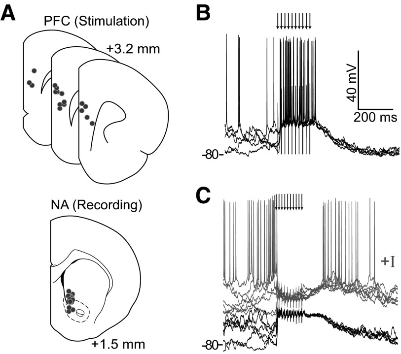
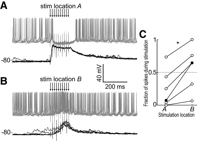
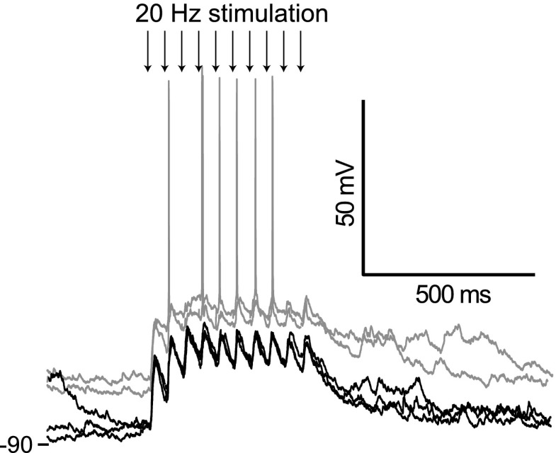
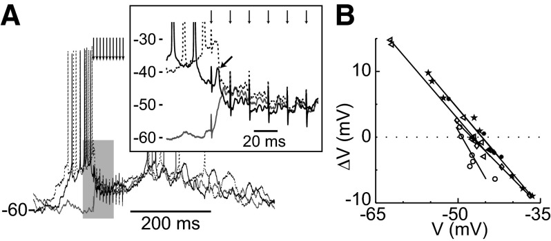
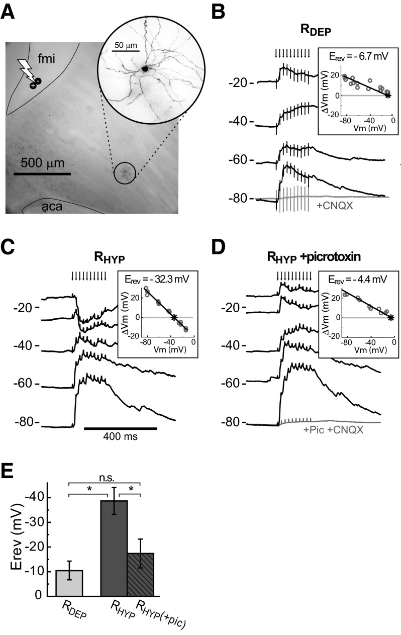
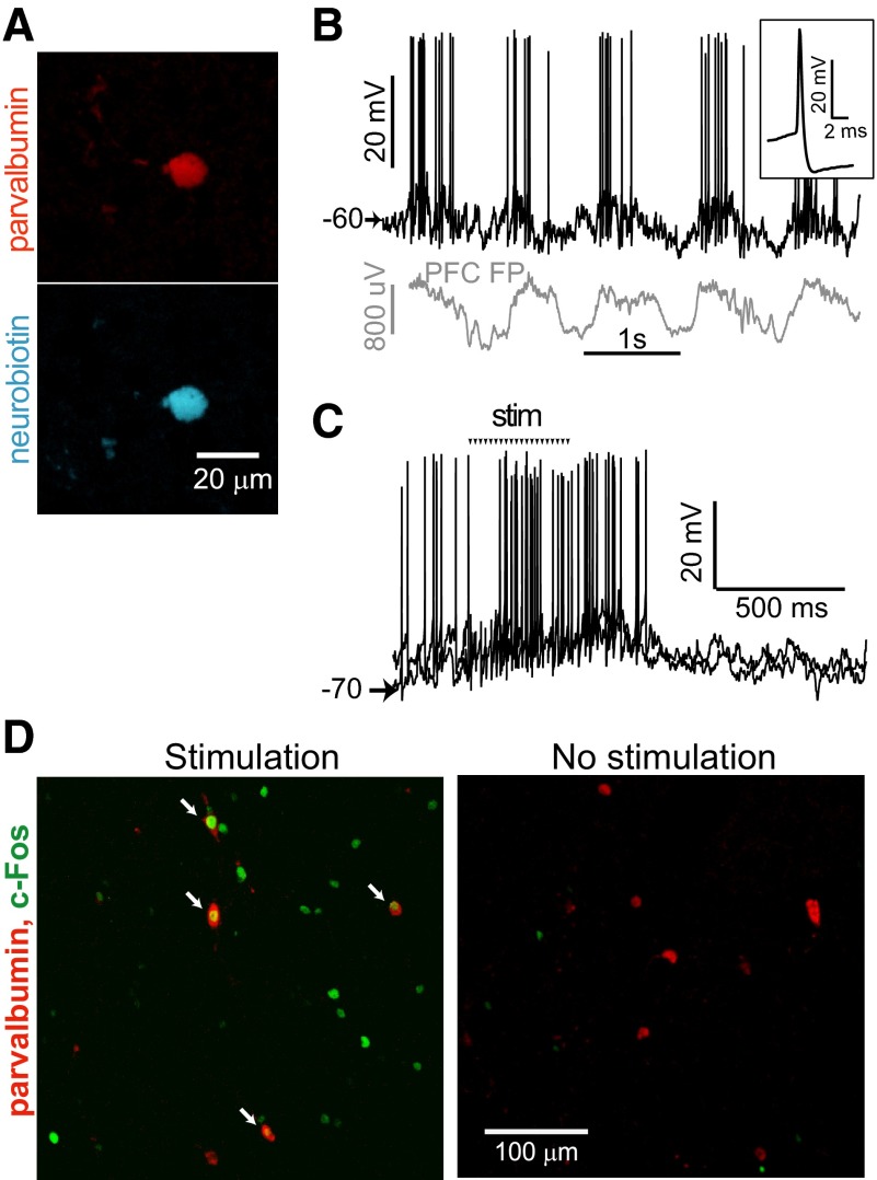
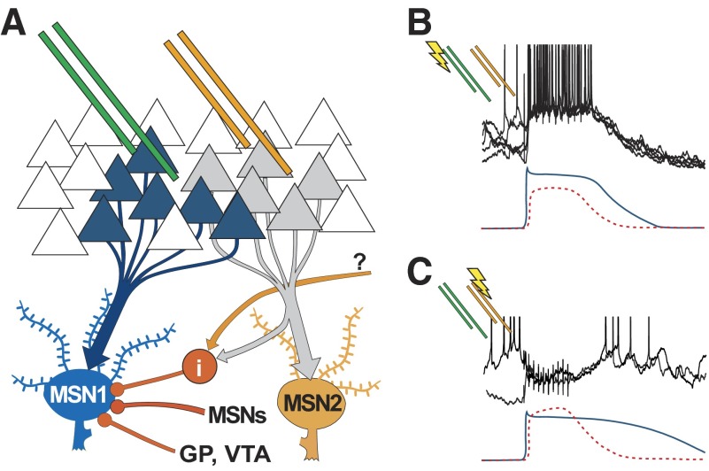
Basal ganglia circuits are organized as parallel loops that have been proposed to compete in a winner-take-all fashion to determine the appropriate behavioral outcome. However, limited experimental support for strong lateral inhibition mechanisms within striatal regions questions this model. Here, stimulation of the prefrontal cortex (PFC) using naturally occurring bursty patterns inhibited firing in most nucleus accumbens (NA) projection neurons. When an excitatory response was observed for one stimulation site, neighboring PFC sites evoked inhibition in the same neuron. Furthermore, PFC stimulation activated interneurons, and PFC-evoked inhibition was blocked by GABA(A) antagonists in corticoaccumbens slice preparations. Thus bursting PFC activity recruits local inhibition in the NA, shaping responses of projection neurons with a topographical arrangement that allows inhibition among parallel corticoaccumbens channels. The data indicate a high order of information processing within striatal circuits that should be considered in models of basal ganglia function and disease.


Figures

FIG. 1.
FIG. 1.
In vivo intracellular recordings of prefrontal cortex (PFC)-evoked nucleus accumbens (NA) medium spiny neuron (MSN) responses. A: illustration of stimulating and recording locations shown as dots. Some neurons were dorsal to the classic NA boundary but within the region receiving afferents from the medial PFC (Voorn et al. 2004). B: overlay of voltage traces from a neuron showing a bursting response evoked by PFC train stimulation. Arrows indicate stimulation pulses here and in subsequent figures. C: overlay of traces from a different neuron showing depolarization and lack of firing with undisturbed membrane potential (dark traces), and a suppression of firing (gray traces) evoked by PFC train stimulation when the neuron was depolarized with constant current. The overlays in B and C include traces recorded with stimuli occurring during up and down states, revealing the independence of responses on the membrane potential state at the time of stimulation.
FIG. 2.
FIG. 2.
Individual neurons can show different responses to 2 adjacent cortical sites. A and B: evoked response of an MSN showing a suppression of firing for stimulation in 1 PFC location (A) but an enhancement of firing for stimulation in a different PFC location (B). Black traces were recorded without current injection; gray traces were recorded with constant intracellular current injection that caused spontaneous firing. C: ratio of firing during stimulation (from 1st pulse to 200 ms later) to firing during and after stimulation (from 1st pulse to 400 ms later) for 6 neurons showing that response pattern depends on stimulation location.
FIG. 3.
FIG. 3.
Corticoaccumbens excitatory postsynaptic potentials (EPSPs) are observed throughout the burst stimulation. Overlay of traces showing responses in a NA neuron to PFC burst stimulation at a lower frequency (20 Hz), showing EPSPs in response to every stimulus in the train (arrows point to the stimuli). The gray traces were obtained from the same neuron when depolarized by intrasomatic current injection, revealing action potential firing evoked by some stimuli.
FIG. 4.
FIG. 4.
Reversal of evoked in vivo responses. A: overlay of traces from a representative cell showing responses to PFC train stimulation under constant artificial depolarization by current injection. Variation in membrane potential prior to stimulation is due to spontaneous activity. An expanded view (inset) of the gray shaded region shows an early component with a highly depolarized reversal (arrow) followed by a convergence of the response at an intermediate potential. B: linear regression (lines) of evoked reversals for inhibited cells (symbols) shows a mean reversal of −47.1 ± 1.5 mV.
FIG. 5.
FIG. 5.
Cortically activated inhibition in cortico-accumbens slices. A: photomontage of a corticoaccumbens slice and a Neurobiotin-stained neuron (inset) illustrating the relative placement of stimulating electrode (bolt symbol) and recorded neuron with respect to the forceps minor (fmi) and anterior commissure (aca). B: overlay of voltage traces in current clamp from a MSN showing depolarization evoked by train stimulation of corticostriatal afferents and attenuation of evoked responses by bath application of 6-cyano-7-nitroquinoxalene-2,3-dione (CNQX) (50 μM; bottom trace). Current was injected to depolarize the neuron to explore the reversal potential (Erev) of the evoked response, which was –6.7 mV as determined by linear regression (inset). Neurons with such depolarized reversals are classified as RDEP. C: overlay of voltage traces from an RHYP MSN that showed a more hyperpolarized reversal of the evoked response at –32.3 mV. D: overlay of voltage traces from the same neuron as in C during bath application of the GABA antagonist picrotoxin (10 μm), which shifted the reversal of the evoked response to a less negative value. E: the addition of picrotoxin shifts the reversal of RHYP neurons to potentials similar to those observed in RDEP neurons.
FIG. 6.
FIG. 6.
Activation of NA interneurons by PFC train stimulation in vivo. A: parvalbumin (red) and Neurobiotin (blue) co-labeling of a fast spiking interneuron recorded intracellularly in vivo in the NA. B: recording from the neuron in a (dark trace) and a simultaneously recorded PFC field potential (PFC FP, gray trace—the polarity was inverted to make it easier to visualize the synchronization) showing spontaneous fluctuations. Inset shows the action potential waveform. C: overlay of traces from the same neuron in response to PFC stimulation. Arrows show timing of the electrical pulses, and stimulus artifacts have been removed for clarity. D: confocal image of a NA section labeled for c-Fos (green) and parvalbumin (red) following in vivo train stimulation (left). The presence of co-labeling (arrows) indicates that PFC train stimulation can activate NA interneurons. A control section from an unstimulated brain (right) shows very little c-Fos immunoreactivity. Scale is the same in both panels.
FIG. 7.
FIG. 7.
Model of cortico-accumbens processing. A: diagram illustrating the hypothetical organization of cortico-accumbens projections in which electrode placement can activate cortical units directly projecting to 1 MSN (MSN1; blue) from 1 position (green pair) while another placement could activate a different cortico-NA channel (orange electrodes and MSN2), driving lateral inhibition via interneurons (i— red) and potentially also via collaterals from other MSNs. Other inputs to the NA could also activate interneurons (question mark). B and C: illustrations of the magnitude and time course of hypothetical excitatory (solid) and inhibitory (dotted) somatic components are shown under previously shown data traces. MSN1 receives a predominantly excitatory input with stimulation via the left (green) electrodes (B) and produces a bursting response. Stimulation via the right (orange) electrodes (C) activates feedforward and feedback GABAergic pathways, resulting in a strong shunting inhibitory input to MSN1 during the stimulation.

References

    1. Alexander GE, Crutcher MD, DeLong MR. Basal ganglia-thalamocortical circuits: parallel substrates for motor, oculomotor, “prefrontal” and “limbic” functions. Prog Brain Res 85: 119–146, 1990. - PubMed
    1. Bartos M, Vida I, Jonas P. Synaptic mechanisms of synchronized gamma oscillations in inhibitory interneuron networks. Nat Rev Neurosci 8: 45–56, 2007. - PubMed
    1. Bennett BD, Bolam JP. Synaptic input and output of parvalbumin-immunoreactive neurons in the neostriatum of the rat. Neuroscience 62: 707–719, 1994. - PubMed
    1. Berke JD Uncoordinated firing rate changes of striatal fast-spiking interneurons during behavioral task performance. J Neurosci 28: 10075–10080, 2008. - PMC - PubMed
    1. Blackwell KT, Czubayko U, Plenz D. Quantitative estimate of synaptic inputs to striatal neurons during up and down states in vitro. J Neurosci 23: 9123–9132, 2003. - PMC - PubMed

Publication types

MeSH terms

LinkOut - more resources