. 2009 Apr;20(7):1903-15.
doi: 10.1091/mbc.e08-09-0950. Epub 2009 Jan 28.

Cooperative assembly and misfolding of CFTR domains in vivo

Affiliations

Cooperative assembly and misfolding of CFTR domains in vivo

Kai Du et al. Mol Biol Cell. 2009 Apr.

Abstract

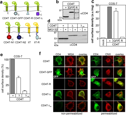
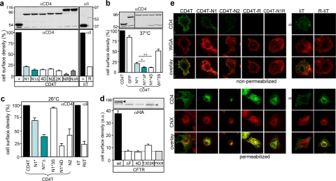
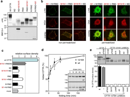
The cystic fibrosis transmembrane conductance regulator (CFTR) architecture consists of two membrane spanning domains (MSD1 and -2), two nucleotide binding domains (NBD1 and -2), and a regulatory (R) domain. Several point mutations lead to the channel misprocessing, with limited structural perturbation of the mutant domain. To gain more insight into the basis of CFTR folding defect, the contribution of domain-wise and cooperative domain folding was assessed by determining 1) the minimal domain combination that is recognized as native and can efficiently escape the endoplasmic reticulum (ER) retention and 2) the impact of mutation on the conformational coupling among domains. One-, two-, three-, and most of the four-domain assemblies were retained at the ER. Solubilization mutations, however, rescued the NBD1 processing defect conceivably by thermodynamic stabilization. The smallest folding unit that traversed the secretory pathway was composed of MSD1-NBD1-R-MSD2 as a linear or split polypeptide. Cystic fibrosis-causing missense mutations in the MSD1, NBD1, MSD2, and NBD2 caused conformational defect in multiple domains. We propose that cooperative posttranslational folding is required for domain stabilization and provides a plausible explanation for the global misfolding caused by point mutations dispersed along the full-length CFTR.


Figures

Figure 1.
Figure 1.
Biosynthetic processing of split CFTR in mammalian cells. (a) The indicated C-terminal CFTR domains (M2N2, RM2N2, N1RM2N2, *RM2N2, and *N1RM2N2) in the absence or presence of complementing N-terminal fragments (M1N1R, M1N1, M1, M1N1*, and M1*, respectively) were transiently expressed in COS7 cells and analyzed by immunoblotting using anti-HA and MM13-4 Abs, recognizing the 3HA epitope in the MSD2 (M2) and the N-terminal tail of the MSD1, respectively. Dotted lines indicate the complex-glycosylated bands, based on endoglycosidase analysis shown in b. (b) Endoglycosidase sensitivity of M2-containing CFTR fragments. Cell lysates, obtained from COS7 cells expressing the indicated CFTR fragments were incubated with endoglycosidase-H (H) or PNGase F (F) and analyzed by immunoblotting with anti-HA antibody. (c) Cell surface density of the CFTR-3HA C-terminal fragments was measured by the immunoperoxidase assay using anti-HA Ab as described in c. Ab binding was normalized for cellular protein (means ± SEM, n = 2–4). (d) Processing of the C-terminal CFTR fragments was monitored by immunostaining in nonpermeabilized (left) and permeabilized (right) cells in the absence or presence of the complementing N-terminal half. The plasma membrane and the ER were visualized by Alexa594-conjugated WGA and CNX staining, respectively. Bar, 10 μm.
Figure 2.
Figure 2.
Cell-based assay to monitor the conformation of membrane tethered soluble CFTR domains. (a) Schematic structure of chimeras consisting of the truncated CD4 (CD4T) or invariant chain (IiT). CD4T-λ and CD4T-λm contain the wt or the mutant (L57C/L69G) λ repressor, respectively. The CD4T-GFP, CD4T-N1, CD4T-N2, and CD4T-R harbor the EGFP, NBD1, NBD2, and R domain, respectively. (b) The indicated CD4T chimeras were transiently expressed in COS7 cells. Equal amounts of proteins were visualized by immunoblotting with anti-CD4 Ab. (c) Cell surface density of the indicated CD4T variants was measured by the immunoperoxidase assay, using primary anti-CD4 Ab and HRP-conjugated secondary Ab in live cells (see Materials and Methods). Amplex-Red fluorescence was normalized for cellular protein and expressed in arbitrary unit (a.u.). Means ± SEM, n ≥ 8. (d) Steady-state expression level of the CD4T chimeras was monitored by immunoblotting following proteasomes inhibition by MG132 (20 μM for 3 h) as indicated. (e) Cell surface expression of CD4T-λand CD4T-λm was measured by the immunoperoxidase assay (means ± SEM, n = 6). (f) Subcellular localization of CD4 chimera was detected by immunostaining with anti-CD4 Ab and Cy2-conjugated secondary Ab in transiently transfected COS7 cells. CD4-GFP was visualized by the EGFP fluorescence. The ER was visualized by anti-calnexin Ab (CNX) and rhodamine-conjugated secondary Ab in permeabilized cells (right). Plasma membrane was stained with WGA in nonpermeabilized cells. Single optical sections, obtained by fluorescence laser confocal microscopy are shown. Bar, 10 μm.
Figure 3.
Figure 3.
Biosynthetic processing of the chimeric CFTR cytosolic domains. (a) Immunoblot and cell surface detection of CD4 and Ii chimeras. The reporter molecules (CD4T and IiT) and the chimeras were transiently expressed in COS7 cells. Immunoblot and cell surface density analysis were performed with anti-CD4 (top left) and anti-Ii (top right) Abs. Cell surface density was expressed as percentage of CD4T or IiT after normalization of the fluorescence signal for cellular proteins. Abbreviations: N1ΔF, NBD1ΔF508; N14D, NBD1(L570D/L571D/I601D/L602D); N2K, NBD2(N1303K); N1R, NBD1-R; N1ΔR, ΔF508-NBD1-R (also see Supplemental Table S1). Means ± SEM, n ≥ 6. (b) Immunoblot and cell surface expression analysis of CD4 chimeras containing NBD1 with boundaries of the crystallized domain (N1*). The N1*ΔF and N1*4D contains the same mutations as defined in a. N1*3S incorporates the F429S, F494N, and Q637R solubilization mutations. Cell surface density and immunoblot analysis were performed as defined in a. Means ± SEM, n ≥ 5. Unpaired, two-tailed t test was used for comparisons. (*p = 0.0006, **p = 0.003; n = 11–15). (c) Cell surface expression of the CD4T and IiT chimeras was rescued at reduced temperature. The cell surface density of the indicated chimeras were measured as described in b, after culturing the cells for 24 h at 26°C. Means ± SEM, n = 3. (d) Expression and cell surface density of wt and mutant CFTR variants in stably transfected BHK cells, determined by immunoblotting and Ab binding assay, respectively. The core- and complex-glycosylated CFTR are depicted by empty and filled arrowhead, respectively. Means ± SEM, n = 3. (e) Indirect immunolocalization of CD4 and Ii chimeras in COS7 cells. Immunostaining of the chimeras with Alexa 594-WGA were performed in nonpermeabilized cells (top). Intracellular staining of the chimeras and CNX was established in permeabilized cells (bottom) as described in Figure 2f. Bar, 10 μm.
Figure 4.
Figure 4.
Intracellular retention of the MSD1 and the MSD2. The MSD1 (M1), EGFP-M1, and MSD2 (M2-3HA) were transiently expressed in COS7 cells. Immunoblotting (a) and cell surface density measurements (b) of MSD1, EGFP-MSD1, and MSD2-3HA (means ± SEM, n = 3). (c) Immunolocalization were performed as described in Figure 3e. Bar, 10 μm.
Figure 5.
Figure 5.
Expression and characterization of CFTR four-domain combinations. CFTR constructs lacking the indicated domain (ΔNBD1, ΔR, and ΔMSD2) (a) and ΔNBD2 (b) were expressed as M1 + RM2N2, M1N1 + M2N2, M1N1R + CD4T-N2, and M1N1RM2 (CFTR-1218X), respectively, in COS7 cells. Equal amounts of cell lysates were immunoblotted (top). Split wt CFTR was expressed as control, demonstrating the electrophoretic mobility shift of the complex-glycosylated R2M2N2 and M2N2 (dotted line) in the presence of complementing N-terminal fragments. The cell surface density of the M2 containing CFTR fragments, wt CFTR, and CD4T-N2 was measured by the immunoperoxidase assay (bottom). Means ± SEM, n = 3. (c) CFTR-1218X is complex-glycosylated. Cell lysates were treated with the endoglycosidase H or Endo F. Immunoblotting was performed with anti-HA Ab. (d) Maturation efficiency of CFTR-1218X was measured by metabolic pulse-chase experiments in stably transfected BHK cells. After a 10-min pulse labeling, cells were chased for 3 h, and CFTR was immunoprecipitated and visualized by fluorography. (e) Metabolic stability of the complex-glycosylated CFTR-1218X. Pulse-labeled BHK cells were chased as indicated, and labeled CFTR was visualized by fluorography and quantified by phosphorimage analysis by using the ImageQuant software (right). (f) In situ protease susceptibility of the wt and C-terminally truncated CFTR. Microsomes from wt, 1218X-, and 1158X CFTR-expressing BHK cells were subjected to limited proteolysis at the indicated trypsin concentration for 15 min on ice. Digestion patterns of CFTR were visualized by immunoblotting with MSD1-, NBD1-, and MSD2-specific anti-CFTR monoclonal Abs.
Figure 6.
Figure 6.
Biosynthetic processing and the folding kinetics of the CFTR-1218X variants. (a) Processing of the split CFTR-1218X. Complex-glycosylation of M2 containing fragments of CFTR-1218X was restored in the presence of complementary N-terminal fragments in transiently transfected COS7 cells. The C- and N-terminal CFTR fragments were visualized by immunoblotting using the anti-HA and MM13-4 Ab, respectively. Dotted line indicates the complex-glycosylated form. (b) Immunolocalization of the M2 containing fragments in cells expressing the indicated N- and C-terminal fragments of the CFTR1218X. Colocalization with Alexa594-WGA and CNX, plasma membrane and ER markers, respectively was performed as described in Figure 1d. Bar, 10 μm. (c) Cell surface density of the M2 containing fragments of COS7 cells transiently expressing the C- and N-terminal fragments of split CFTR-1218X. The immunoperoxidase assay was performed with anti-HA Ab. Wt CFTR and its split variant (M1N1 + RM2N2) were used as positive controls. Means ± SEM, n = 3. (d) Folding kinetics of wt and 1218X CFTR. Stably transfected BHK cells were pulse labeled for 8 min with [35S]methionine and [35S]cysteine and chased in BFA (10 μg/ml) containing medium for 3 h (inset). Folding was terminated by depleting the cellular ATP content as described in Materials and Methods. CFTR was immunoprecipitated and visualized by fluorography (inset). The CFTR radioactivity was quantified by phosphorimage analysis. Similar results were obtained in the absence of BFA. Data are means (±SEM, n = 5) and expressed as percentage of the maximum amount of radioactivity associated with CFTR. (e) Missense mutations provoke the intracellular retention of the CFTR-1218X. COS7 cells were transiently transfected with the indicated construct and biosynthetic processing was visualized by the accumulation of the complex-glycosylated form (arrowhead) with immunoblotting (top). All the mutants remained core-glycosylated. Cell surface density of CFTR-1218X variants was measured by anti-HA Ab binding assay as described in Figure 2c (bottom).
Figure 7.
Figure 7.
Global and domain-wise conformational perturbations of CFTR variants harboring missense mutations. (a) Schematic localization of CF associated point mutations in CFTR used in this study. (b) Point mutations prevent the biosynthetic processing of CFTR detected by immunoblotting in stably transfected BHK cells. Equal amounts of cell lysates were probed with anti-HA Ab. The BFA-induced accumulation of the folded, core-glycosylated wt CFTR in cells exposed to BFA for 24 h is illustrated by immunoblotting (bottom right). (c–g) Comparison of the in situ trypsin susceptibility of wild type and mutant CFTRs. Isolated microsomes expressing the indicated CFTR variant were digested at the indicated concentration of trypsin as described in Figure 6f. CFTR proteolytic fragments were visualized by immunoblotting with the following antibodies: anti-N-MSD1 (MM13-4; c), anti-NBD1 (L12B4; d), anti-NBD1 (660; e); anti-HA (MSD2 specific; f), and anti-NBD2 (M3A7; g). The proteolytic degradation intermediate representing the MSD1-, NBD1-, MSD2- and NBD2-containing fragment, based on multiple epitope localization, molecular masses and ATP binding are indicated by rectangles (Du et al., 2005; Cui et al., 2007). Note that in f, proteolytic digestion was performed on microsomes isolated from BFA-treated wt CFTR-expressing BHK cell to facilitate comparison of the core-glycosylated wt MSD2 with its mutant counterparts.

References

    1. Anelli T., Sitia R. Protein quality control in the early secretory pathway. EMBO J. 2008;27:315–327. - PMC - PubMed
    1. Ashman J. B., Miller J. A role for the transmembrane domain in the trimerization of the MHC class II-associated invariant chain. J. Immunol. 1999;163:2704–2712. - PubMed
    1. Baker J. M., Hudson R. P., Kanelis V., Choy W. Y., Thibodeau P. H., Thomas P. J., Forman-Kay J. D. CFTR regulatory region interacts with NBD1 predominantly via multiple transient helices. Nat. Struct. Mol. Biol. 2007;14:738–745. - PMC - PubMed
    1. Barriere H., Nemes C., Du K., Lukacs G. L. Plasticity of polyubiquitin recognition as lysosomal targeting signals by the endosomal sorting machinery. Mol. Biol. Cell. 2007;18:3952–3965. - PMC - PubMed
    1. Barriere H., Nemes C., Lechardeur D., Khan-Mohammad M., Fruh K., Lukacs G. L. Molecular basis of oligoubiquitin-dependent internalization of membrane proteins in mammalian cells. Traffic. 2006;7:282–297. - PubMed

Publication types

MeSH terms

Substances