. 2009 Apr;296(4):C801-10.
doi: 10.1152/ajpcell.00620.2008. Epub 2009 Jan 28.

Polyamines regulate E-cadherin transcription through c-Myc modulating intestinal epithelial barrier function

Affiliations

Polyamines regulate E-cadherin transcription through c-Myc modulating intestinal epithelial barrier function

Lan Liu et al. Am J Physiol Cell Physiol. 2009 Apr.

Abstract

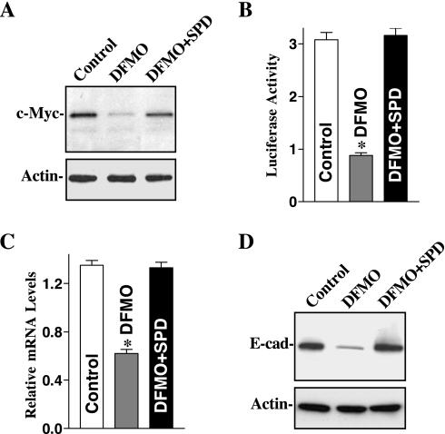
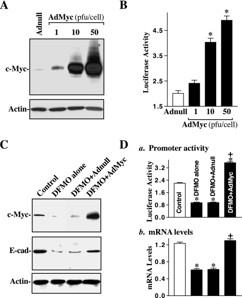
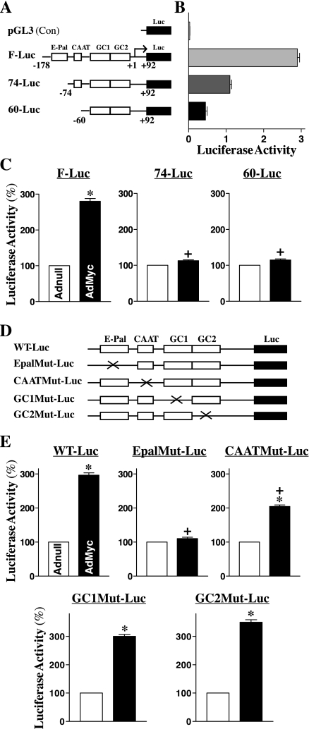
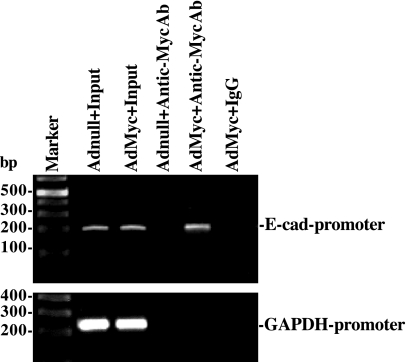
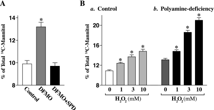
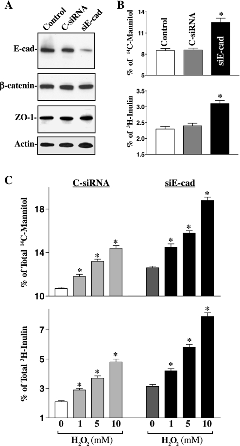
The integrity of the intestinal epithelial barrier depends on intercellular junctions that are highly regulated by numerous extracellular and intracellular factors. E-cadherin is found primarily at the adherens junctions in the intestinal mucosa and mediates strong cell-cell contacts that have a functional role in forming and regulating the epithelial barrier. Polyamines are necessary for E-cadherin expression, but the exact mechanism underlying polyamines remains elusive. The current study was performed to determine whether polyamines induce E-cadherin expression through the transcription factor c-Myc and whether polyamine-regulated E-cadherin plays a role in maintenance of the epithelial barrier integrity. Decreasing cellular polyamines reduced c-Myc and repressed E-cadherin transcription as indicated by a decrease in levels of E-cadherin promoter activity and its mRNA. Forced expression of the c-myc gene by infection with adenoviral vector containing c-Myc cDNA stimulated E-cadherin promoter activity and increased E-cadherin mRNA and protein levels in polyamine-deficient cells. Experiments using different E-cadherin promoter mutants revealed that induction of E-cadherin transcription by c-Myc was mediated through the E-Pal box located at the proximal region of the E-cadherin promoter. Decreased levels of E-cadherin in polyamine-deficient cells marginally increased basal levels of paracellular permeability but, remarkably, potentiated H(2)O(2)-induced epithelial barrier dysfunction. E-cadherin silencing by transfection with its specific small interfering RNA also increased vulnerability of the epithelial barrier to H(2)O(2). These results indicate that polyamines enhance E-cadherin transcription by activating c-Myc, thus promoting function of the epithelial barrier.


Figures

Fig. 1.
Fig. 1.
Effect of depletion of cellular polyamines by inhibiting ornithine decarboxylase (ODC) on c-Myc and E-cadherin expression in intestinal epithelial cell (IEC)-6 cells. Cells were grown in control cultures, cultures in which ODC activity was inhibited by 5 mM α-difluoromethylornithine (DFMO), and cultures inhibited with DFMO and supplemented with 5 μM spermidine (SPD) for 6 days. A: representative immunoblots. Whole cell lysates were harvested, applied to each lane (20 μg) equally, and subjected to electrophoresis on a 10% acrylamide gel. Levels of c-Myc protein were identified with a specific antibody that recognizes c-Myc. After the membrane was stripped, it was reprobed with actin that served as an internal control for equal loading. B: E-cadherin promoter activity in cells described in A. Cells were grown in medium containing either DFMO alone or DFMO + SPD for 4 days and were then transfected using the E-cadherin promoter luciferase reporter construct. After incubation for 48 h in the same culture conditions, transfected cells were harvested and assayed for luciferase activity. Data were normalized according to β-galactosidase activity from cotransfected plasmid pRSV β-galactosidase. Values are means ± SE of data from 3 separate experiments. *P < 0.05 compared with controls and cells exposed to DFMO + SPD. C: levels of E-cadherin mRNA in cells described in A. Total cellular RNA was isolated, and levels of E-cadherin mRNA were measured using real-time quantitative PCR analysis. Values are means ± SE of data from 3 separate experiments. *P < 0.05 compared with control and cells treated with DFMO + SPD. D: representative immunoblots of E-cadherin in cells described in A. E-cad, E-cadherin. Data are representative of 3 independent experiments showing similar results.
Fig. 2.
Fig. 2.
Effect of c-Myc overexpression on E-cadherin expression in normal and polyamine-deficient cells. A: representative immunoblots. The c-Myc adenoviral expression vector (AdMyc) was constructed in which the complete open reading frame of the human c-Myc cDNA was cloned to pShuttle of the Adeno-X expression system under the control of the human cytomegalovirus immediate-early promoter pCMV. Cells were infected with the AdMyc or adenoviral vector containing no c-Myc cDNA (Adnull) at a multiplicity of infection of 1–50 plaque-forming units (pfu)/cell, and c-Myc expression was analyzed 48 h after the infection. B: changes in E-cadherin promoter activity in cells described in A. The luciferase activity was measured 48 h after the transfection with the E-cadherin-promoter luciferase reporter construct, and data were normalized according to β-galactosidase activity from cotransfected plasmid pRSV β-galactosidase. Values are means ± SE of data from 3 separate experiments. *P < 0.05 compared with cells infected with the Adnull. C: effect of ectopic expression of c-Myc on E-cadherin expression in polyamine-deficient cells. Cells were grown in control cultures and cultures containing 5 mM DFMO for 4 days and then infected with the AdMyc or Adnull at a concentration of 10 pfu/cell. After 48 h in the presence of DFMO, whole cell lysates were harvested; levels of c-Myc and E-cadherin proteins were measured by Western blot analysis. Equal loading was monitored by actin immunoblotting. D: changes in levels of E-cadherin promoter activity (a) and its mRNA (b) in cells described in C. Values are means ± SE of data from 3 separate results. *P < 0.05 compared with controls. +P < 0.05 compared with DFMO-treated cells and cells treated with DFMO and then infected with the Adnull.
Fig. 3.
Fig. 3.
Effect of ectopic expression of the c-myc gene on E-cadherin promoter activity after mutation of different binding sites. A: schematic representation of deletion of E-cadherin promoter luciferase (Luc) reporter constructs. E-Pal, CAAT, GC1, and GC2 indicate the relative locations of specific binding sites within the E-cadherin-promoter. B: basal activity of various deletion mutants of the E-cadherin promoter in IEC-6 cells without c-Myc overexpression. Cells were transfected with different deletion mutants of E-cadherin-promoter, and levels of the luciferase reporter activity were detected 48 h after the transfection. Results were normalized relative to β-galactosidase activity from cotransfected plasmid pRSV β-galactosidase. Values are means ± SE of data from 3 separate experiments. C: changes in luciferase reporter activity of deletion constructs after c-Myc overexpression. Cells were infected with either the AdMyc or Adnull at s concentration of 10 pfu/cell for 24 h and then transfected using different E-cadherin promoter luciferase reporter deletion constructs. The levels of luciferase activity was assayed 48 h after transfection. Values are means ± SE of data from 3 separate experiments. *P < 0.05 compared with cells transfected with the Adnull. +P < 0.05 compared with cells infected with the AdMyc and then transfected with the F-Luc. D: schematic representation of various point mutants. Mutagenic oligonucleotides were designed to hybridize to the E-cadherin-promoter construct to create the mutant (Mut) where the E-Pal, CAAT, GC1, or GC2 consensus site was eliminated by making 2 base changes. E: levels of reporter gene activity. Values are means ± SE of data from 3 separate experiments. *P < 0.05 compared with cells infected with the Adnull. +P < 0.05 compared with cells infected with the AdMyc and then transfected with the WT-Luc.
Fig. 4.
Fig. 4.
Activity of c-Myc binding to E-cadherin promoter as measured by chromatin immunoprecipitation (ChIP) analysis. The association of c-Myc with the E-cadherin promoter (between −144 and 61) was determined using an anti-c-Myc antibody (Ab); IgG was used as a negative control. Cross-linked chromatin isolated from cells infected with either the AdMyc or Adnull was immunoprecipitated using an anti-c-Myc Ab, and the associated chromosomal DNA fragments were amplified by PCR using E-cadherin promoter-specific primers and GAPDH promoter-specific primers as described in materials and methods. The expected size of the PCR product was ∼205 bp. Chromosomal DNA input was subject to the same procedures and served as a positive control. Three experiments were performed that showed similar results.
Fig. 5.
Fig. 5.
Changes in paracellular permeability in control and polyamine-deficient IEC-6 cells with or without challenge with H2O2. A: basal levels of paracellular permeability. After cells were grown in control cultures or cultures containing DFMO or DFMO + SPD for 4 days, they then were trypsinized, plated at confluent density on the insert, and maintained under the same culture conditions for an additional 48 h. Membrane-impermeable tracer molecule, [14C]mannitol, was added to the insert medium, and the entire basal medium was collected 2 h thereafter for paracellular tracer flux assays. Values are means ± SE of data from 6 samples. *P < 0.05 compared with control cells and cells treated with DFMO + SPD. B: paracellular permeability in controls (a) and polyamine-deficient cells (b) after challenge with different concentrations of H2O2 for 3 h. After cells were treated with H2O2 for 1 h, [14C]mannitol was added to the insert medium, and levels of [14C]mannitol were measured 2 h thereafter. Values are means ± SE of data from 6 samples. *P < 0.05 compared with control cells.
Fig. 6.
Fig. 6.
Effect of E-cadherin silencing on basal levels of paracellular permeability. A: representative immunoblots for E-cadherin, β-catenin, and zonula occludens-1 (ZO-1) proteins. Cells were transfected with specific small interfering RNA (siRNA) targeting the coding region of E-cadherin mRNA (siE-cad) or control siRNA (C-siRNA) for 48 h, and levels of E-cadherin, β-catenin, and ZO-1 proteins were measured by Western blot analysis. B: paracellular permeability in cells described in A. After cells were transfected with siE-cad or C-siRNA, they were plated at confluent density on the insert and maintained for an additional 48 h. Levels of paracellular permeability were measured 2 h after addition of [14C]mannitol or [3H]inulin. Values are means ± SE of data from 6 samples. *P < 0.05 compared with controls and cells transfected with C-siRNA. C: changes in levels of paracellular permeability after challenge with H2O2 in E-cadherin knockdown cells described in A. Cells were transfected with siE-cad or C-siRNA for 48 h, and levels of paracellular permeability were measured 2 h after addition of [14C]mannitol or [3H]inulin. Values are means ± SE of data from 6 samples. *P < 0.05 compared with cells transfected with C-siRNA.

Similar articles

Cited by

References

    1. Angst BD, Marcozzi C, Magee AI. The cadherin superfamily. J Cell Sci 114: 625–626, 2001. - PubMed
    1. Angst BD, Marcozzi C, Magee AI. The cadherin superfamily: diversity in form and function. J Cell Sci 114: 629–641, 2001. - PubMed
    1. Balda MS, Whitney JA, Flores C, González S, Cereijido M, Matter K. Functional dissociation of paracellular permeability and transepithelial electrical resistance and disruption of the apical-basolateral intramembrane diffusion barrier by expression of a mutant tight junction membrane protein. J Cell Biol 134: 1031–1049, 1996. - PMC - PubMed
    1. Batsché E, Muchardt C, Behrens J, Hurst HC, Crémisi C. RB and c-Myc activate expression of the E-cadherin gene in epithelial cells through interaction with transcription factor AP-2. Mol Cell Biol 18: 3647–3658, 1998. - PMC - PubMed
    1. Behrens J, Löwrick O, Klein-Hitpass L, Birchmeier W. The E-cadherin promoter: functional analysis of a GC-rich region and an epithelial cell-specific palindromic regulatory element. Proc Natl Acad Sci USA 88: 11495–11499, 1991. - PMC - PubMed

Publication types

MeSH terms

LinkOut - more resources