. 2009 Jan 28;29(4):1140-51.
doi: 10.1523/JNEUROSCI.3073-08.2009.

The contribution of synaptic inputs to sustained depolarizations in reticulospinal neurons

Affiliations

The contribution of synaptic inputs to sustained depolarizations in reticulospinal neurons

Myriam Antri et al. J Neurosci. .

Abstract

Sensory stimulation elicits sustained depolarizations in lamprey reticulospinal (RS) cells for which intrinsic properties were shown to play a crucial role. The depolarizations last up to minutes, and we tested whether the intrinsic properties required the cooperation of synaptic inputs to maintain RS cells depolarized for such long periods of time. Ascending spinal inputs to RS cells were reversibly blocked by applying xylocaine over the rostral spinal cord segments. The duration of the sustained depolarizations was markedly reduced. The membrane potential oscillations in tune with locomotor activity that were present under control condition were also abolished. The contribution of excitatory glutamatergic inputs was then assessed by applying CNQX and AP-5 over one of two simultaneously recorded homologous RS cells on each side of the brainstem. The level of sensory-evoked depolarization decreased significantly in the cell exposed to the antagonists compared with the other RS cell monitored as a control. In contrast, local application of glycine only produced a transient membrane potential hyperpolarization with a marked reduction in the amplitude of membrane potential oscillations. Locally applied strychnine did not change the duration of the sustained depolarizations, suggesting that mechanisms other than glycinergic inhibition are involved in ending the sustained depolarizations in RS cells. It is concluded that excitatory glutamatergic inputs, including ascending spinal feedback, cooperate with intrinsic properties of RS cells to maintain the cells depolarized for prolonged periods, sustaining long bouts of escape swimming.


Figures
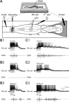
Figure 1.
Figure 1.
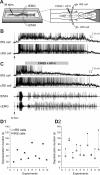
Effects of blocking spinal cord feedback with xylocaine on sensory-evoked sustained depolarizations. A, The experimental set-up. Stimulation of the ascending lateral columns (S stim) was used to confirm that xylocaine blocked the connections between spinal cord and brainstem. Sustained depolarizations were intracellularly recorded from RS cells after mechanical pressure (M stim) applied to the skin covering the head, or electrical stimulation of the trigeminal nerve (Vth stim). Xylocaine was locally ejected in a circular bath tightly fitted on the rostralmost spinal segments. B, C, Sustained depolarizations of different durations (top) and EMG activity (bottom) recorded in two different preparations under control (B1 and C1), xylocaine (B2 and C2) and after washout (B3 and C3). The EMG activity was recorded at segmental level 30. Note the absence of EMG activity under xylocaine (B2 and C2) and the recovery of EMG bursts after washout (B3 and C3).
Figure 2.
Figure 2.
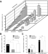
Durations of sensory-evoked depolarizations before, during xylocaine and after washout. A, Plot showing the mean depolarization duration (±SEM) of 8–10 trials measured in 20 experiments under control (ascending order), xylocaine, and after washout obtained in 10 experiments. Note the thick black line separating the experiments showing no significant decrease in duration after xylocaine from those that did. B, Histogram illustrating the mean depolarization durations for the 20 experiments shown in A, under control (black), xylocaine (gray) and after washout (white). C, Histogram depicting the mean depolarization durations under the three experimental conditions, for all the trials having, in control condition, a depolarization duration shorter (left), and longer (right) than 25 s. **p < 0.01; ***p < 0.001; ns, Nonsignificant.
Figure 3.
Figure 3.
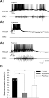
Effects of blocking ascending spinal inputs with xylocaine on sensory-evoked depolarizations in the absence of sensory feedback from the dorsal columns. A, After the bilateral lesion of the dorsal columns, sustained depolarizations were intracellularly recorded from RS cells in response to electrical stimulation of the trigeminal nerve (Vth stim). Sustained depolarizations (top) and EMG activity (bottom) recorded under control (A1), after xylocaine locally ejected in a circular bath fitted on the rostralmost spinal segments (A2) and after washout (A3), in one preparation. Note that EMG bursts recorded at segmental level 30 were abolished under xylocaine (A2, EMG) and recovered after washout (A3, EMG). B, Histogram of the mean depolarization durations of five experiments under the three experimental conditions. **p < 0.01, t test.
Figure 4.
Figure 4.
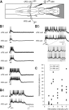
Sensory-evoked depolarizations in two simultaneously recorded homologous RS cells. A, Drawing illustrating the experimental set-up. Paired intracellular recordings from two homologous RS cells located on either side of the brain were performed. One cell always served as the control RS cell (cRS cell) allowing drug ejections to be made over the tRS cell on the opposite side. B1–B5, Sustained depolarization recorded simultaneously in both cells at different trigeminal nerve stimulation intensities (7.0, 7.6, 11.0, 8.0 and 8.0 μA, respectively). The depolarization varied similarly in the two RS cells from very short (B1, 3 s), to much longer (B5, 50 s). Note the alternating pattern of membrane potential oscillations observed in B5 (inset). C, Plot of the stimulus-response relationship for the control (cRS cell, black circles) and the test (tRS cell, gray circles) RS cells illustrated in B. The stimulation intensity varied from 7 to 11 μA and the response durations from 3 to 55 s.
Figure 5.
Figure 5.
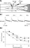
Effects of a local application of a mixture of CNQX (1 mm) and AP-5 (2 mm) on sensory-evoked subthreshold responses in a RS cell. A, Experimental set-up whereby glutamate receptor antagonists were locally pressure ejected over one RS cell. B1, A typical gradual reduction of the trigeminal-induced EPSPs is shown for one RS cell in control and after 20-, 30-, and 60-s exposure to the glutamate receptor antagonists. B2, Time course of EPSP areas (relative to control value) in four RS cells (different preparations) after exposure to the glutamate receptor antagonists. Traces in B1 are from the experiment represented by the inverted triangles.
Figure 6.
Figure 6.
Effects of a local application of a mixture of CNQX (1 mm) and AP-5 (2 mm) over one of two homologous RS cells. A, The experimental set-up. B, Paired intracellular recordings from two homologous RS cells on either side of the brainstem (control and test RS cells) showing sustained depolarizations induced by mechanical stimulation of the tail (arrow) under control condition. C, Local ejections of the glutamate receptor antagonists (gray horizontal bar) over the tRS cell. Note that the membrane potential depolarization decreased in the cell exposed to the glutamate receptor antagonists without affecting the contralateral control neuron (cRS cell). EMG activity (bottom traces) recorded at segmental level 30 from the control (cEMG) and test (tEMG) sides. Recordings in B and C are from the same neurons. D, Duration of sensory-evoked sustained depolarizations for the control (cRS cell, black circles) and the test (tRS cell, gray circles) RS cells before (D1) and after local ejections of CNQX and AP-5 over the test RS cells (D2). Note that in the 10 pairs of simultaneously recorded homologous RS cells, the control RS cells and test RS cells display sustained depolarizations with similar durations under control condition (D1). After a local ejection of CNQX and AP-5, the sustained depolarizations in the test RS cells are in general of a shorter duration (D2).
Figure 7.
Figure 7.
Effect of the glutamate receptor antagonists, CNQX and AP-5, on the amplitude of RS cell membrane potential oscillations during locomotion. A, Gradual decrease in the oscillation amplitude of one RS cell after the local application of CNQX (1 mm) and AP-5 (2 mm) at time = 0 s (dotted line). B, Plot showing the mean amplitude (±SEM) of a series of eight consecutive membrane potential oscillations measured in control and 40 s after the local ejections of CNQX and AP-5 for five RS cells.
Figure 8.
Figure 8.
Effects of local ejections of glycine (1 mm) on sensory-evoked sustained depolarizations, membrane potential oscillations, and input resistance of RS cells. A, Local application of glycine (gray horizontal bar) over one (tRS cell) of two homologous cells produce a transient hyperpolarization without ending the sustained depolarization evoked by a mechanical stimulation (M stim) of the tail. The EMG activity (bottom traces) was recorded at segmental level 30 from both sides (cEMG, tEMG). B, Local ejections of glycine also strongly reduce the membrane potential oscillatory pattern, without affecting the swimming episode (cEMG and tEMG recordings). C, Effect of bath-applied glycine (100 μm) on the membrane input resistance. Responses to current injections in presence of TTX before and after glycine ejection (top traces). The V–I relationship (bottom) is plotted before and after glycine ejection.
Figure 9.
Figure 9.
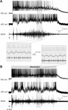
Effects of local ejections of strychnine (500 μm) on sensory-evoked sustained depolarizations. A, The control condition. Sustained depolarizations intracellularly recorded from two homologous RS cells, control (cRS) and test (tRS), in response to mechanical tail stimulation (M stim, arrow). Swimming activity was recorded ipsilateral to the tRS cell at segmental level 30 (tEMG). B, Effect of strychnine. The drug was locally applied over the test RS cell (tRS). Note that strychnine was applied from the beginning to the end of the sustained depolarization (gray horizontal bar). Insets, Parts of the traces (shaded areas) recorded under control or strychnine are shown at a faster time scale below (A) or above (B) the respective recordings. Traces in A and B are from the same pair of neurons. Note that strychnine neither prolonged the sustained depolarization of the tRS cell nor affected the cRS cell on the opposite side. Strychnine had also no effect on the swimming bout. However, strychnine abolished the membrane potential oscillations and increased the firing frequency of the tRS cell.
Figure 10.
Figure 10.
Schematic illustration of the proposed cellular cascade of events involved in sensory-evoked sustained depolarizations in lamprey RS cells. Top left and bottom, Glutamate released (green circles) from trigeminal sensory relay cells (triggering synaptic inputs) that project onto RS cells first activates AMPA and NMDA receptors, producing membrane depolarization and allowing calcium entry (black circles). Intracellular calcium then activates an ICAN that generates a sustained depolarization in RS cells. Excitatory synaptic inputs (maintaining synaptic inputs), would provide additional excitation during the sustained depolarizations. Inset top right, Schematic illustration of proposed cooperation between synaptic inputs and intrinsic membrane properties in RS cells. Triggering synaptic inputs (hatched filling) provide the initial excitation. Intrinsic membrane properties (gray filling) would then provide most of the following excitation to keep the cell depolarized. The superimposed maintaining synaptic inputs (black filling) would generate membrane potential oscillations as well as provide the additional excitation to maintain the RS cells depolarized for long periods of time as the intrinsic excitation would start to die-off.

Similar articles

Cited by

References

    1. Alford S, Dubuc R. Glutamate metabotropic receptor mediated depression of synaptic inputs to lamprey reticulospinal neurones. Brain Res. 1993;605:175–179. - PubMed
    1. Baufreton J, Garret M, Rivera A, de la Calle A, Gonon F, Dufy B, Bioulac B, Taupignon A. D5 (not D1) dopamine receptors potentiate burst-firing in neurons of the subthalamic nucleus by modulating an l-type calcium conductance. J Neurosci. 2003;23:816–825. - PMC - PubMed
    1. Brocard F, Dubuc R. Differential contribution of reticulospinal cells to the control of locomotion induced by the mesencephalic locomotor region. J Neurophysiol. 2003;90:1714–1727. - PubMed
    1. Buchanan JT. Identification of interneurons with contralateral, caudal axons in the lamprey spinal cord: synaptic interactions and morphology. J Neurophysiol. 1982;47:961–975. - PubMed
    1. Buchanan JT. The roles of spinal interneurons and motoneurons in the lamprey locomotor network. Prog Brain Res. 1999;123:311–321. - PubMed

Publication types

MeSH terms

LinkOut - more resources