. 2009 Jan 28;29(4):1202-11.
doi: 10.1523/JNEUROSCI.4516-08.2009.

Fgfr1 is required for cortical regeneration and repair after perinatal hypoxia

Affiliations

Fgfr1 is required for cortical regeneration and repair after perinatal hypoxia

Devon M Fagel et al. J Neurosci. .

Abstract

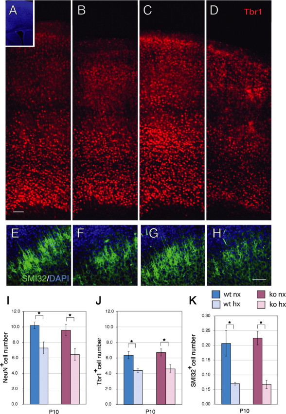
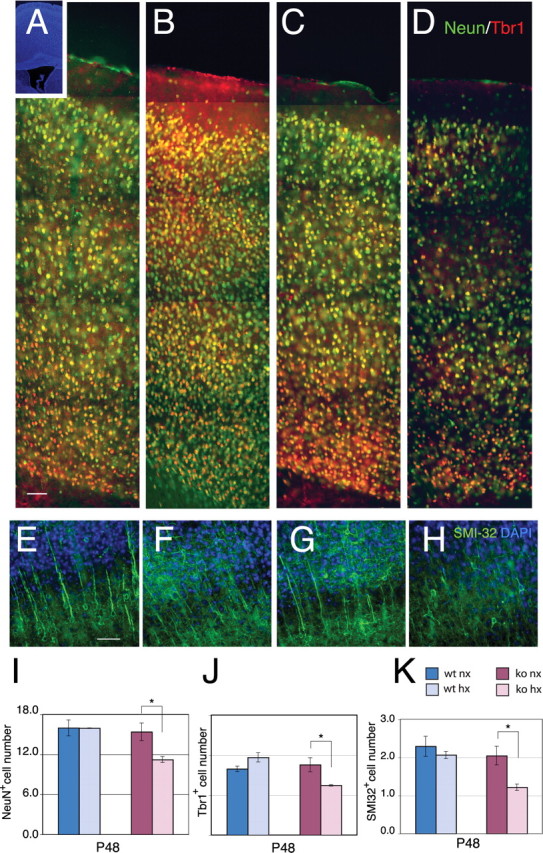
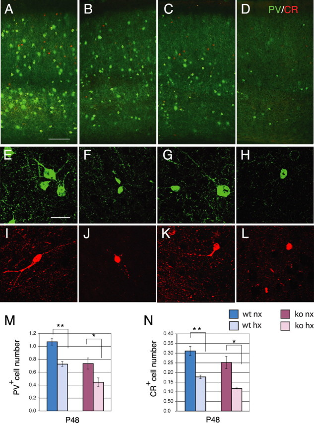
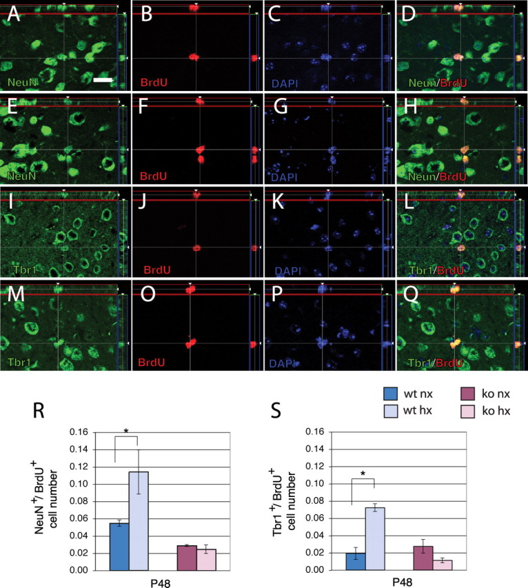
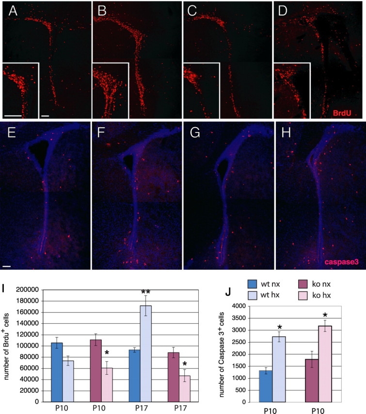
Chronic postnatal hypoxia causes an apparent loss of cortical neurons that is reversed during recovery (Fagel et al., 2006). The cellular and molecular mechanisms underlying this plasticity are not understood. Here, we show that chronic hypoxia from postnatal days 3 (P3) to 10 causes a 30% decrease in cortical neurons and a 24% decrease in cortical volume. T-brain-1 (Tbr1)(+) and SMI-32(+) excitatory neuron numbers were completely recovered 1 month after the insult, but the mice showed a residual deficit in Parvalbumin(+) and Calretinin(+) GABAergic interneurons. In contrast, hypoxic mice carrying a disrupted fibroblast growth factor receptor-1 (Fgfr1) gene in GFAP+ cells [Fgfr1 conditional knock-out (cKO)], demonstrated a persistent loss of excitatory cortical neurons and a worsening of the interneuron defect. Labeling proliferating progenitors at P17 revealed increased generation of cortical NeuN(+) and Tbr1(+) excitatory neurons in wild-type mice subjected to hypoxic insult, whereas Fgfr1 cKO failed to mount a cortical neurogenetic response. Hypoxic wild-type mice also demonstrated a twofold increase in cell proliferation in the subventricular zone (SVZ) at P17 and a threefold increase in neurogenesis in the olfactory bulb (OB) at P48, compared with normoxic mice. In contrast, Fgfr1 cKO mice had decreased SVZ cell proliferation and curtailed reactive neurogenesis in the OB. Thus, the activation of FGFR-1 in GFAP+ cells is required for neuronal recovery after neonatal hypoxic injury, which is attributable in part to enhanced cortical and OB neurogenesis. In contrast, there is incomplete recovery of inhibitory neurons after injury, which may account for persistent behavioral deficits.


Figures

Figure 1.
Figure 1.
Cortical neuron number and thickness are decreased after exposure to hypoxia. A–D, Immunostaining for Tbr1 (red) in the cerebral cortex of wild-type (A, B) or Fgfr1 cKO mice (C, D) at P10 under normoxia (A, C) or after hypoxia from P3 to P10 (B, D). Inset shows DAPI low magnification. Each panel is the composite of four 20× images demonstrating the entire extent of the cerebral cortex. E–H, SMI-32 (green) and DAPI (blue) stained cortices of wild-type (E, F) or Fgfr1 cKO mice (G, H) at P10 under normoxia (E, G) or after hypoxia from P3 to P10 (F, H). Scale bars, 100 μm. I–K, Total number of NeuN (I), Tbr1 (J) and SMI-32 (K) immunoreactive neurons in the cerebral cortex by stereological analyses in wild-type (blue bars) and Fgfr1 cKO (red bars) mice. Values are expressed in 106 units. N = 3 for each group. *p < 0.05 by ANOVA with Sheffe post hoc test.
Figure 2.
Figure 2.
Neuron numbers recover in hypoxic wild-type mice but not in Fgfr1 cKO mice. A–D, Double immunostaining for NeuN (green) and Tbr1 (red) in the P48 cerebral cortex of wild-type (A, B) or Fgfr1 cKO mice (C, D) under normoxia (A, C) or after hypoxia (B, D). Inset shows DAPI low magnification. Each panel is the composite of five 20× images. E–H, SMI-32- (green) and DAPI- (blue) stained cortices of wild-type (E, F) or Fgfr1 cKO mice (G, H) at P48 under normoxia (E, G) or after hypoxia from P3 to P10 (F, H). Scale bars, 100 μm. I–K, Total number of NeuN (I), Tbr1 (J) and SMI-32 (K) immunoreactive neurons in the cerebral cortex by stereological analyses in wild-type (blue bars) and Fgfr1 cKO (red bars) mice. Values are expressed in 106 units. N = 3 for each group. *p < 0.05 by ANOVA with Sheffe post hoc test.
Figure 3.
Figure 3.
Cortical interneuron number is decreased at P48 in both hypoxic wild-type and Fgfr1 cKO mice. A–D, Double immunostaining for PV (green) and CR (red) in the P48 cerebral cortex of wild-type (A, B) or Fgfr1 cKO mice (C, D) under normoxia (A, C) or after hypoxia (B, D). Each panel is the composite of two images to show all layers of the cerebral cortex. E–L, Individual PV+ and CR+ neurons in hypoxia-reared mice (F, J, H, L) and their normoxic counterparts (E, I, G, K). Scale bars: (A–D) 100 μm; (E–L) 10 μm. M, N, Total number of PV (M) and CR (N) immunoreactive neurons by stereological analyses in wild-type (blue bars) and Fgfr1 cKO (red bars) mice. Values are expressed in 106 units. N = 3 for each group. *p < 0.05 and **p < 0.01 by ANOVA with Sheffe post hoc test.
Figure 4.
Figure 4.
Newly generated NeuN+ and Tbr1+ cortical neurons are increased at P48 in hypoxic wild-type but not in Fgfr1 cKO mice. A–Q, Apotome 1 μm single slices of BrdU staining in the P48 cerebral cortex double-immunostained with NeuN (A–H) or Tbr1 (I–Q). BrdU was injected at P17 and analysis performed at P48. The image analyses on the z-axis are shown in the side panels. NeuN (A, E), Tbr1 (I, M), BrdU (B, F, J, O), DAPI (C, G, K, P), and merged images (D, H, L, Q). BrdU colocalization is observed in neurons born at P17 in normoxic (A–D, I–L) and hypoxic cortex (E–H, M–Q). R, S, Total number of NeuN/Brdu (R) and Tbr1/Brdu (S) double-labeled neurons in the cerebral cortex by stereological analyses in wild-type (blue bars) and Fgfr1 cKO (red bars). Scale bar, 10 μm. Values are expressed in 106 units. N = 3 for each group. *p < 0.05 by ANOVA with Sheffe post hoc test.
Figure 5.
Figure 5.
Increase in proliferation in the SVZ during recovery requires Fgfr1. A–D, BrdU immunostaining in the SVZ at P17 of wild-type (A, B) or Fgfr1 cKO mice (C, D) under normoxia (A, C) or after hypoxia (B, D). Insets show high magnifications. E–H, Caspase-3 immunostaining in the SVZ at P10 of wild-type (E, F) or Fgfr1 cKO (G, H) mice under normoxia (E, G) or after hypoxia (F, H). I, J, Total number of BrdU+ cells (I) at P10 and P17 and caspase-3+ cells (J) at P10 in the SVZ by stereological analyses in wild-type (blue bars) and Fgfr1 cKO (red bars). Scale bar, 100 μm. N = 3 for each group. *p < 0.05 and **p < 0.01 by ANOVA with Sheffe post hoc test.

References

    1. Aguirre A, Gallo V. Postnatal neurogenesis and gliogenesis in the olfactory bulb from NG2-expressing progenitors of the subventricular zone. J Neurosci. 2004;24:10530–10541. - PMC - PubMed
    1. Allin M, Henderson M, Suckling J, Nosarti C, Rushe T, Fearon P, Stewart AL, Bullmore ET, Rifkin L, Murray R. Effects of very low birthweight on brain structure in adulthood. Dev Med Child Neurol. 2004;46:46–53. - PubMed
    1. Alvarez-Buylla A, Garcia-Verdugo JM. Neurogenesis in adult subventricular zone. J Neurosci. 2002;22:629–634. - PMC - PubMed
    1. Androutsellis-Theotokis A, Leker RR, Soldner F, Hoeppner DJ, Ravin R, Poser SW, Rueger MA, Bae SK, Kittappa R, McKay RD. Notch signalling regulates stem cell numbers in vitro and in vivo. Nature. 2006;442:823–826. - PubMed
    1. Arvidsson A, Collin T, Kirik D, Kokaia Z, Lindvall O. Neuronal replacement from endogenous precursors in the adult brain after stroke. Nat Med. 2002;8:963–970. - PubMed

Publication types

MeSH terms