. 2009 Jun;34(7):1764-73.
doi: 10.1038/npp.2008.234. Epub 2009 Jan 28.

Enhanced sensitivity of the MRL/MpJ mouse to the neuroplastic and behavioral effects of chronic antidepressant treatments

Affiliations

Enhanced sensitivity of the MRL/MpJ mouse to the neuroplastic and behavioral effects of chronic antidepressant treatments

Darrick T Balu et al. Neuropsychopharmacology. 2009 Jun.

Abstract

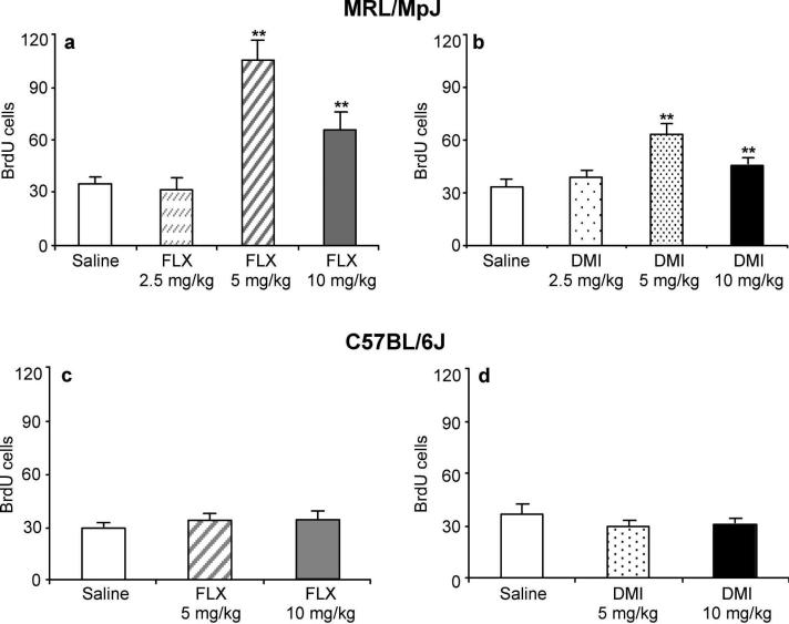
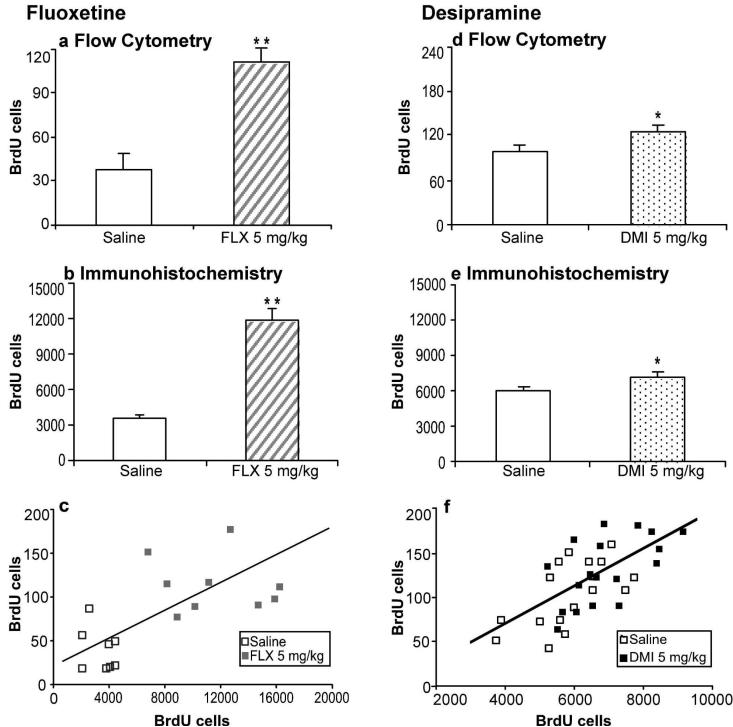
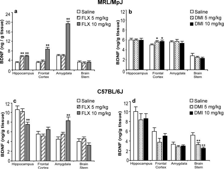
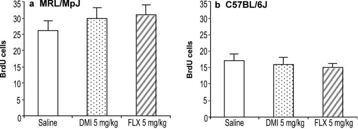
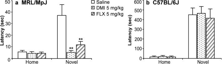
Chronic administration of antidepressant drugs produce changes in neuroplasticity and behavior in rodents, effects that may be associated with the slow emergence of clinical therapeutic effects. Owing to the uncertainty over the effects of chronic antidepressant treatments in mice, these experiments compared the regulation of neurogenesis, neurotrophin levels, and behavior produced by chronic antidepressant treatments between two inbred mouse strains, MRL/MpJ and C57BL/6J. The MRL/MpJ strain is associated with enhanced wound healing and tissue regeneration, whereas C57BL/6J mice are used commonly for behavioral studies. Proliferation and survival of hippocampal progenitor cells were measured using flow cytometry, a new platform that rapidly quantifies the incorporation of 5-bromo-2-deoxyuridine (BrdU). Hippocampal cell proliferation was increased significantly after chronic administration of fluoxetine (FLX: 5, 10 mg/kg, intraperitoneal (i.p.), b.i.d.) or desipramine (DMI: 5, 10 mg/kg, i.p., b.i.d.) for 21 days in MRL/MpJ mice, but not in C57BL/6J mice. Hippocampal progenitor cells born prior to chronic antidepressant treatments were not affected in either mouse strain. Protein levels of brain-derived neurotrophic factor (BDNF) in MRL/MpJ mice were elevated significantly in the frontal cortex, hippocampus, and amygdala after chronic FLX treatment, but increased only in the frontal cortex by chronic DMI. In contrast, BDNF levels in C57BL/6J mice were decreased in the hippocampus and increased in the amygdala after chronic FLX, and were decreased in the brain stem after chronic DMI. Novelty-induced hypophagia (NIH) was used to examine a behavioral effect produced by chronic antidepressant treatment. MRL/MpJ mice, chronically administered FLX or DMI, had significantly shorter latencies to consume food when exposed to a novel environment than untreated mice, whereas there were no effects on the behavior of C57BL/6J mice. In conclusion, robust effects of chronic antidepressant treatments on hippocampal cell proliferation and BDNF levels paralleled the ability of these drugs to produce changes in NIH behavior in MRL/MpJ, while none of these effects were produced in C57BL/6J mice. The greater responsiveness of MRL/MpJ mice may be important for drug discovery, for genetic studies, and for understanding the neural mechanisms underlying the physiological and behavioral effects of chronic antidepressant treatments.


Figures

Figure 1
Figure 1
Chronic fluoxetine and desipramine treatments elevated cell proliferation in MRL/MpJ mice but not C57BL/6J mice. (a) MRL/MpJ mice were administered saline (n = 15) or fluoxetine (FLX; 2.5, 5, 10 mg/kg b.i.d.; n = 10/group) for 21 days. The 5 and 10 mg/kg doses of FLX increased cell proliferation (F(3,38) = 21.89, P < 0.0001) (b) MRL/MpJ mice were administered saline (n = 15) or desipramine (DMI; 2.5, 5, 10 mg/kg b.i.d.; n = 10/group) for 21 days. DMI (5 and 10 mg/kg) significantly increased cell proliferation (F(3,38) = 8.77, P < 0.001). (c) C57BL/6J mice were administered saline (n = 10) or fluoxetine (FLX; 5 or 10 mg/kg b.i.d.; n = 10/group) for 21 days. FLX did not alter cell proliferation (F(2,21) = 0.60, P = 0.56). (d) C57BL/6J mice were administered saline (n = 10) or desipramine (DMI; 5 or 10 mg/kg b.i.d.; n = 10/group) for 21 days. DMI did not increase cell proliferation (F(2,22) = 1.17, P = 0.32). Values are expressed as the number of BrdU positive cells per 10,000 7-AAD events. Bars represent mean values + 1 s.e.m. Asterisks (**) indicate groups that differed significantly from control (P < 0.005) according to Dunnett's test.
Figure 2
Figure 2
Comparison of BrdU incorporation in the hippocampus measured by flow cytometry and immunohistochemistry following chronic antidepressant treatments. One cohort of MRL/MpJ mice (a-c) was administered saline (n = 9; open bars) or fluoxetine (FLX; 5 mg/kg b.i.d.; n = 9; striped bars), while another (d-f) was administered saline (n = 17; open bars) or desipramine (DMI; 5 mg/kg b.i.d.; n = 18; dotted bars) for 21 days. (a, d) Cell proliferation was measured in one hippocampal lobe by flow cytometry. Values are expressed as the number of BrdU positive cells per 10,000 7-AAD events. FLX (t(18) = 5.94, P < 0.001) and DMI (t(33) = 2.12, P = 0.04) significantly increased cell proliferation. (b, e) The contralateral hemisphere was sectioned and BrdU-labeled cells in the hippocampus were counted using immunohistochemistry. Values are expressed as the number of BrdU positive cells per hippocampus. FLX (t(15) = 6.47, P < 0.001) and DMI (t(33) = 2.66, P = 0.01) significantly increased cell proliferation. Bars represent mean values + 1 s.e.m. Asterisks indicate groups that differed significantly from saline (* P < 0.05, ** P < 0.005), according to unpaired Student's t-test. (c, f) Scatter plots showing the correlation between results obtained from individual mice using flow cytometry and immunohistochemistry (FLX: r(15) = 0.62, P = 0.007; (r(33) = .65, P < 0.001).
Figure 3
Figure 3
Differential regulation of BDNF protein levels by chronic fluoxetine and desipramine treatments in MRL/MpJ mice and C57BL/6J mice. (a) MRL/MpJ and (c) C57BL/6J mice were administered saline (open bar, n = 10) or fluoxetine (FLX, n = 10/group) at 5 (striped bar) or 10 mg/kg (grey bar) b.i.d. for 21 days. In MRL/MpJ mice, FLX increased BDNF levels in the hippocampus (F(2,25) = 52.62, P < 0.001), frontal cortex (F(2,25) = 44.81 P < 0.001), and amygdala (F(2,25) = 155.08, P < 0.001). In C57BL/6J mice, FLX decreased BDNF levels significantly in the hippocampus (F(2,25) = 7.40, P < 0.003) and increased BDNF levels in the amygdala (F(2,25) = 14.04, P < 0.001). All other regional comparisons were not statistically significant. (b) MRL/MpJ and (d) C57BL/6J mice were administered saline (open bars, n = 10) or desipramine (DMI, n = 10/group) at 5 (dotted bars) or 10 (black bar) mg/kg b.i.d. for 21 days. In MRL/MpJ mice, DMI increased BDNF levels only in the frontal cortex (F(2,27) = 4.33, P = 0.02). In C57BL/6J mice, DMI decreased BDNF levels in the brain stem (F(2,27) = 11.36, P < 0.001). All other regional comparisons were not statistically significant. Values are expressed as the amount of BDNF protein per gram of tissue. Bars represent mean values + 1 s.e.m. Asterisks indicate groups that differed significantly from saline treatment (* P < 0.05, ** P < 0.005) according to Dunnett's test.
Figure 4
Figure 4
Chronic treatments with fluoxetine or desipramine did not affect the survival of newly born hippocampal progenitor cells. Cells were labeled by administration of BrdU (100 mg/kg i.p. for 4 days) prior to the initiation of chronic treatment. (a) MRL/MpJ mice or (b) C57BL/6J mice (n = 10 mice/group) were administered saline (open bar), fluoxetine (FLX; 5 mg/kg b.i.d.; striped bar), or desipramine (DMI; 5 mg/kg b.i.d.; dotted bar) for 21 days. Values for ANOVA were not statistically significant (P > 0.05). Values are expressed as the number of BrdU positive cells per 10,000 7-AAD events. Bars represent mean values + 1 s.e.m.
Figure 5
Figure 5
Chronic treatments with fluoxetine and desipramine reduced reactivity to novelty in the NIH test in MRL/MpJ mice but not in C57BL/6J mice. (a) MRL/MpJ mice (n = 7-9) or (b) C57BL/6J mice (n = 7-9) were administered saline (open bar), desipramine (DMI; 5 mg/kg b.i.d.; dotted bar), or fluoxetine (FLX; 5 mg/kg; striped bar) for 21 days. The chronic antidepressant treatments reduced the latency to initiate feeding in a novel environment for MRL/MpJ mice (F(2,25) = 8.13, P = 0.002) but not for C57BL/6J mice (F(2,20) = 0.10, P = .90). The antidepressant treatments did not alter home cage feeding for both strains. Values are expressed as the latency (sec) to consume peanut butter chips in either the home cage (Home) or a novel environment (Novel). Bars represent mean values + 1 s.e.m. Asterisks (**) indicate groups that differed significantly from saline (P < 0.005) according to Dunnett's test.

Similar articles

Cited by

References

    1. Airan RD, Meltzer LA, Roy M, Gong Y, Chen H, Deisseroth K. High-speed imaging reveals neurophysiological links to behavior in an animal model of depression. Science. 2007;317:819–823. - PubMed
    1. Altar CA, Whitehead RE, Chen R, Wortwein G, Madsen TM. Effects of electroconvulsive seizures and antidepressant drugs on brain-derived neurotrophic factor protein in rat brain. Biol Psychiatry. 2003;54:703–709. - PubMed
    1. Baker KL, Daniels SB, Lennington JB, Lardaro T, Czap A, Notti RQ, Cooper O, Isacson O, Frasca S, Jr., Conover JC. Neuroblast protuberances in the subventricular zone of the regenerative MRL/MpJ mouse. J Comp Neurol. 2006;498:747–761. - PubMed
    1. Balu DT, Lucki I. Adult hippocampal neurogenesis: Regulation, functional implications, and contribution to disease pathology. Neurosci Biobehav Rev. 2008 - PMC - PubMed
    1. Balu DT, Hoshaw BA, Malberg JE, Rosenzweig-Lipson S, Schechter LE, Lucki I. Differential regulation of central BDNF protein levels by antidepressant and nonantidepressant drug treatments. Brain Res. 2008;1211:37–43. - PMC - PubMed

Publication types

MeSH terms