

doi: 10.1002/pro.15.
Crystal structures of the X-domains of a Group-1 and a Group-3 coronavirus reveal that ADP-ribose-binding may not be a conserved property
Affiliations
- PMID: 19177346
- PMCID: PMC2708038
- DOI: 10.1002/pro.15
Item in Clipboard
Crystal structures of the X-domains of a Group-1 and a Group-3 coronavirus reveal that ADP-ribose-binding may not be a conserved property
Protein Sci.
2009 Jan.
Abstract
The polyproteins of coronaviruses are cleaved by viral proteases into at least 15 nonstructural proteins (Nsps). Consisting of five domains, Nsp3 is the largest of these (180-210 kDa). Among these domains, the so-called X-domain is believed to act as ADP-ribose-1''-phosphate phosphatase or to bind poly(ADP-ribose). However, here we show that the X-domain of Infectious Bronchitis Virus (strain Beaudette), a Group-3 coronavirus, fails to bind ADP-ribose. This is explained on the basis of the crystal structure of the protein, determined at two different pH values. For comparison, we also describe the crystal structure of the homologous X-domain from Human Coronavirus 229E, a Group-1 coronavirus, which does bind ADP-ribose.
Figures
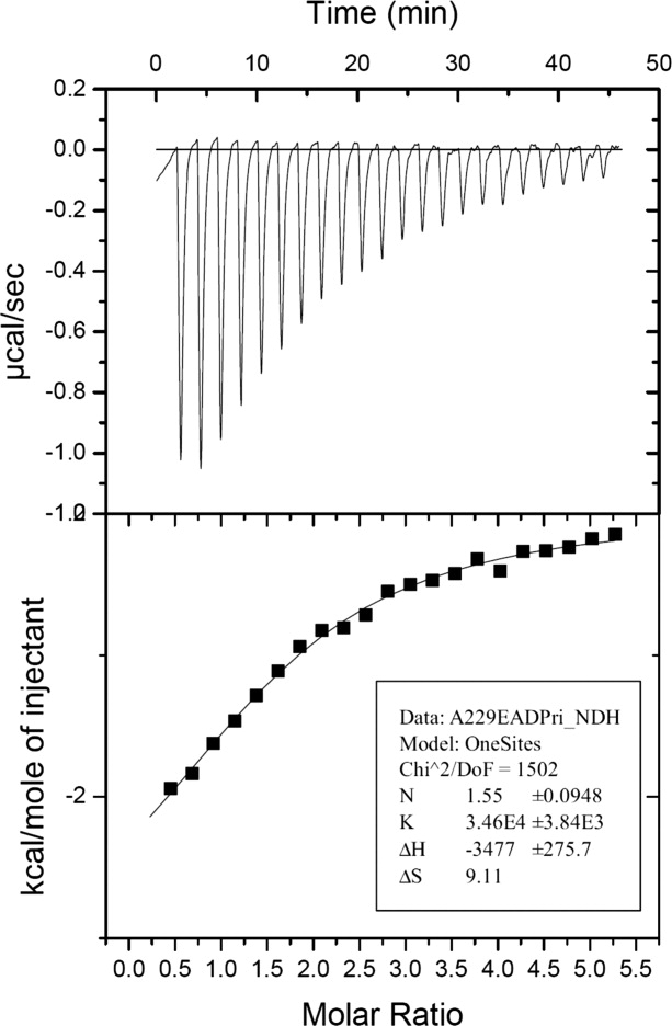
Isothermal titration calorimetry profile for the binding of ADP-ribose to HCoV-229E X-domain.
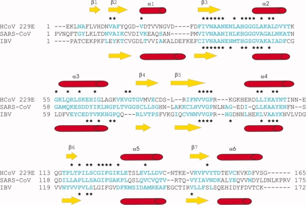
Structure-based sequence alignment of the X-domains of HCoV 229E and IBV (strain Beaudette) with the homologue in SARS-CoV. Secondary structure elements of the HCoV-229E X-domain and the IBV X-domain are represented above and below the alignment, respectively. Amino acid residues labeled in cyan have been included in the calculation of the r.m.s.d. value. Asterisks above and below the alignment indicate aligned and conserved amino acid residues.
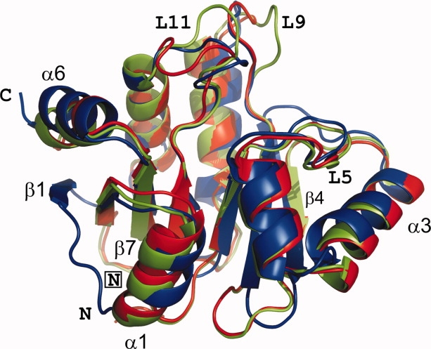
Ribbon-representation of the superimposed X-domains of HCoV 229E (blue) and IBV (strain Beaudette) (green, pH 8.5; red, pH 5.6). N- and C-termini are indicated; the symbol “N” is framed for IBV-X.
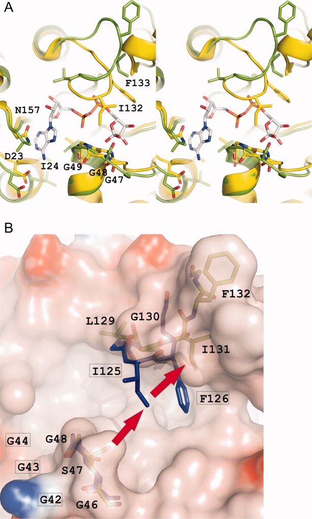
A: Stereo representation showing the binding of ADP-ribose to the X-domain of SARS-CoV, compared with the equivalent site in IBV-X8.5. The ADP-ribose molecule and the amino acid residues involved in the binding are represented as sticks. Carbon atoms of the ADP-ribose are colored white, carbon atoms of the protein are colored yellow and green, for SARS-X and IBV-X, respectively. Nitrogens and oxygens are colored blue and red, respectively. B: Residues 42GGG44 and 122SCGIFG127 (L11) of 229E-X (sticks, blue carbon atoms), likely involved in binding ADP-ribose, superimposed onto the corresponding site (46GSG48 and 128SLGIFG133) in IBV-X8.5 (transparent surface colored according to electrostatic potential, green sticks). Residue labels for 229E-X are framed. Note that Ile 131, and with it, the entire loop L11, in IBV-X is “pushed away” (red arrows) due to steric hindrance from Ser 47, thereby destroying the binding site for ADP-ribose.
Similar articles
-
The SARS-unique domain (SUD) of SARS coronavirus contains two macrodomains that bind G-quadruplexes.PLoS Pathog. 2009 May;5(5):e1000428. doi: 10.1371/journal.ppat.1000428. Epub 2009 May 15. PLoS Pathog. 2009. PMID: 19436709 Free PMC article.
-
Crystal structures of two coronavirus ADP-ribose-1''-monophosphatases and their complexes with ADP-Ribose: a systematic structural analysis of the viral ADRP domain.J Virol. 2009 Jan;83(2):1083-92. doi: 10.1128/JVI.01862-08. Epub 2008 Nov 5. J Virol. 2009. PMID: 18987156 Free PMC article.
-
The ADP-ribose-1''-monophosphatase domains of severe acute respiratory syndrome coronavirus and human coronavirus 229E mediate resistance to antiviral interferon responses.J Gen Virol. 2011 Aug;92(Pt 8):1899-1905. doi: 10.1099/vir.0.031856-0. Epub 2011 Apr 27. J Gen Virol. 2011. PMID: 21525212
-
Players in ADP-ribosylation: Readers and Erasers.Curr Protein Pept Sci. 2016;17(7):654-667. doi: 10.2174/1389203717666160419144846. Curr Protein Pept Sci. 2016. PMID: 27090904 Review.
-
Nsp3 of coronaviruses: Structures and functions of a large multi-domain protein.Antiviral Res. 2018 Jan;149:58-74. doi: 10.1016/j.antiviral.2017.11.001. Epub 2017 Nov 8. Antiviral Res. 2018. PMID: 29128390 Free PMC article. Review.
Cited by
-
The VIZIER project: overview; expectations; and achievements.Antiviral Res. 2010 Aug;87(2):85-94. doi: 10.1016/j.antiviral.2010.02.326. Epub 2010 Mar 10. Antiviral Res. 2010. PMID: 20226212 Free PMC article. Review.
-
Bioinformatics and functional analyses of coronavirus nonstructural proteins involved in the formation of replicative organelles.Antiviral Res. 2016 Nov;135:97-107. doi: 10.1016/j.antiviral.2016.10.005. Epub 2016 Oct 13. Antiviral Res. 2016. PMID: 27743916 Free PMC article. Review.
-
The SARS-unique domain (SUD) of SARS coronavirus contains two macrodomains that bind G-quadruplexes.PLoS Pathog. 2009 May;5(5):e1000428. doi: 10.1371/journal.ppat.1000428. Epub 2009 May 15. PLoS Pathog. 2009. PMID: 19436709 Free PMC article.
-
The macro domain as fusion tag for carrier-driven crystallization.Protein Sci. 2017 Feb;26(2):365-374. doi: 10.1002/pro.3073. Epub 2016 Nov 2. Protein Sci. 2017. PMID: 27774698 Free PMC article.
-
Drug similarity and structure-based screening of medicinal compounds to target macrodomain-I from SARS-CoV-2 to rescue the host immune system: a molecular dynamics study.J Biomol Struct Dyn. 2022 Jan;40(1):523-537. doi: 10.1080/07391102.2020.1815583. Epub 2020 Sep 8. J Biomol Struct Dyn. 2022. PMID: 32897173 Free PMC article.
References
-
- Cavanagh D. Coronavirus avian infectious bronchitis virus. Vet Res. 2007;38:281–297. - PubMed
MeSH terms
Substances
LinkOut - more resources
Full Text Sources
Other Literature Sources
