. 2009 Jan;18(1):134-46.
doi: 10.1002/pro.24.

Backbone structure of a small helical integral membrane protein: A unique structural characterization

Affiliations

Backbone structure of a small helical integral membrane protein: A unique structural characterization

Richard C Page et al. Protein Sci. 2009 Jan.

Abstract

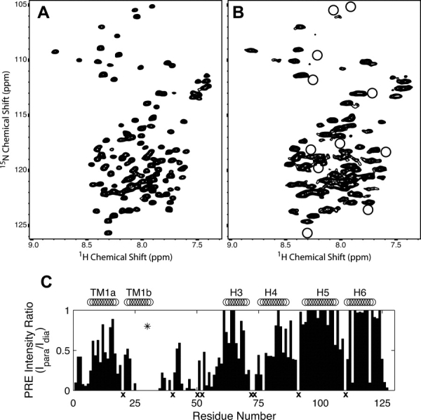
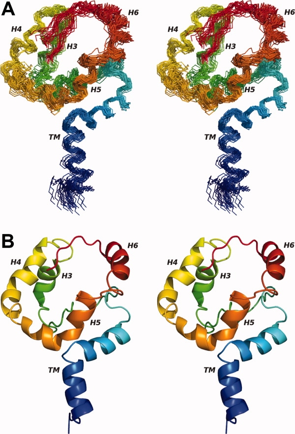
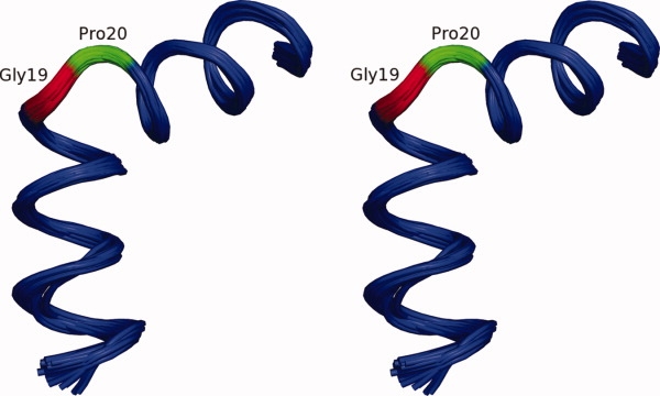
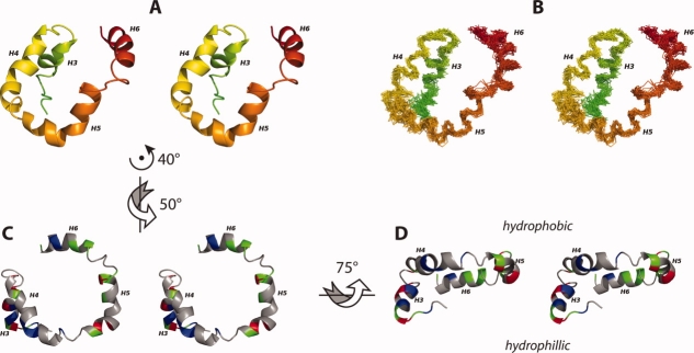
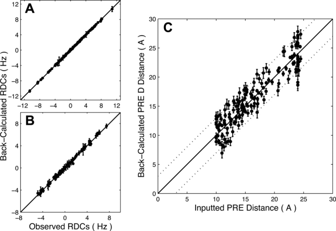
The structural characterization of small integral membrane proteins pose a significant challenge for structural biology because of the multitude of molecular interactions between the protein and its heterogeneous environment. Here, the three-dimensional backbone structure of Rv1761c from Mycobacterium tuberculosis has been characterized using solution NMR spectroscopy and dodecylphosphocholine (DPC) micelles as a membrane mimetic environment. This 127 residue single transmembrane helix protein has a significant (10 kDa) C-terminal extramembranous domain. Five hundred and ninety distance, backbone dihedral, and orientational restraints were employed resulting in a 1.16 A rmsd backbone structure with a transmembrane domain defined at 0.40 A. The structure determination approach utilized residual dipolar coupling orientation data from partially aligned samples, long-range paramagnetic relaxation enhancement derived distances, and dihedral restraints from chemical shift indices to determine the global fold. This structural model of Rv1761c displays some influences by the membrane mimetic illustrating that the structure of these membrane proteins is dictated by a combination of the amino acid sequence and the protein's environment. These results demonstrate both the efficacy of the structural approach and the necessity to consider the biophysical properties of membrane mimetics when interpreting structural data of integral membrane proteins and, in particular, small integral membrane proteins.


Figures

Figure 1
Figure 1
The topology of Rv1761c can be inferred from initial solution NMR data. The Cα chemical shift index (A), residual dipolar couplings in 5.5% (B), and 5% 20-A (C), polyacrylamide gel alignment media and 1H/15N-NOE intensities (D) indicate the presence of a kinked TM helix (TM1a, TM1b) and four extramembrane (EM) helices (H3, H4, H5, and H6). The Rv1761c amino acid sequence (E) is also shown. The positions of the TM domain and helices H3, H4, H5, and H6 are shown through the fitting of dipolar waves to both sets of RDC data. The fitting of the TM domain to two separate waves indicates a kink between segments TM1a and TM1b. 1H/15N-NOE data from the linker region (residues 35–60) indicate varied flexibility; the turn between residues 45–53 undergoes motion similar to the transmembrane helix and Helices H5 and H6 while the loop regions (residues 35–44 and 54–57) before and after the turn display increased dynamics. Residue numbering corresponds to wild-type Rv1761c. Data for the 24 residue amino-terminal fusion tag are not shown.
Figure 2
Figure 2
A comparison of 1H/15N-HSQC spectra of samples with diamagnetic and paramagnetic labels. Spectra acquired for diamagnetic dMTSL-labeled (A) and paramagnetic MTSL-labeled (B) F30C-Rv1761c illustrate resonance broadening due to paramagnetic relaxation enhancement (PRE). Residues in the paramagnetic sample (B) that were broadened so that they were no longer detectable are circled. An intensity plot shows the normalized PRE intensity ratio (Ipara/Idia) used to calculate PRE-derived distances used for Rv1761c structural calculations (C). The location of the spin label at F30C (asterisk) and locations of the TM helix and EM helices H3, H4, H5, and H6 are also shown. Residues for which no 1H/15N resonances were observed are marked with an x (C).
Figure 3
Figure 3
The 30 lowest energy backbone structures (A) are shown in a wide-eyed stereo diagram along with a ribbon diagram of the lowest energy structure (B). The ribbon representation clearly shows the kink of the TM helix and helical propensity of the EM domain. Both diagrams are colored from blue (amino terminus) to red (carboxyl terminus). Structures are aligned to the average backbone structure using all NH, Cα, and C′ atoms. Figure generated using PyMOL.
Figure 4
Figure 4
The kinked TM domain in stereo view was tightly defined within the structure family at a backbone rmsd of 0.40 Å. Despite the 70° kink at the Gly19-Pro20-Gly21-Gly22 sequence, the departure from helical structure is limited to residues Gly19 (red) and Pro20 (green). Structures are aligned to the average backbone structure using the NH, Cα, and C′ atoms for residues 7–30. Figure generated using PyMOL.
Figure 5
Figure 5
The EM domain of Rv1761c is characterized by a set of four helices (A, B). These four helices are organized such that H4 and H5 are co-planar and both H3 and H6 are behind the H4/H5 plane (A, B). The helical domain is shown as a ribbon model (A) and a backbone trace for the 30 lowest energy structures (B) colored in a rainbow (as in Fig. 3A, B). The 30 superimposed backbone structures (B) are aligned to the average backbone structure using NH, Cα, and C′ atoms from helices H3, H4, H5, and H6 (Residues 59–67, 73–86, 90–103, and 110–120). The U-shaped H4/H5 helical pair forms a hydrophobic surface (C) with the amphipathic Helix H6 situated just below the H4/H5 plane and the hydrophilic Helix H3 located well below the H4/H5 plane (D). For C, D positively charged residues are colored red (Arg, His, Lys), negatively charged residues are colored blue (Asp, Glu), nonpolar residues are colored grey (Ala, Gly, Ile, Leu, Met, Pro, Phe, Ser, Thr, Val) and polar uncharged residues are colored green (Asn, Cys, Gln, Trp, Tyr). Throughout this figure, the flexible carboxyl terminal region (residues 122–127) and the flexible linker region between the TM and H3 are not shown. Arrows and axes indicate the approximate rotations used to transform between the orientations depicted in A, C, and D. Figure generated using PyMOL..
Figure 6
Figure 6
Good correlations are seen for observed and back-calculated RDC and PRE data. Correlation coefficients were r = 0.999 for 5.5% (A) and r = 0.997 for 5% 20-A (B) polyacrylamide gel RDC data. Observed RDC data points are the experimental values used for the structure calculation. Back-calculated RDC data points represent the average value of each RDC determined by SVD using Xplor-NIH for the 30 calculated structures. Error bars represent the rmsd for each back-calculated dipolar coupling over the family of 30 structures (error bars for some 5.5% neutral PAG and 5% 20-A PAG data points are smaller than the symbols shown). Average PRE distances calculated from the structure family (back-calculated) agree well with the PRE distance data used for the structural calculations (C). The correlation coefficient for PRE distance data was r = 0.926. Dotted lines indicate ±3 Å error bounds for the PRE distance data. PRE distance error bars represent the rmsd for each back-calculated PRE distance over the family of 30 structures.

References

    1. Korepanova A, Gao FP, Hua Y, Qin H, Nakamoto RK, Cross TA. Cloning and expression of multiple integral membrane proteins from Mycobacterium tuberculosis in Escherichia coli. Protein Sci. 2005;14:148–158. - PMC - PubMed
    1. Gong XM, Franzin CM, Thai K, Yu J, Marassi FM. Nuclear magnetic resonance structural studies of membrane proteins in micelles and bilayers. Methods Mol Biol. 2007;400:515–529. - PMC - PubMed
    1. Stouffer AL, Acharya R, Salom D, Levine AS, Di Costanzo L, Soto CS, Tereshko V, Nanda V, Stayrook S, Degrado WF. Structural basis for the function and inhibition of an influenza virus proton channel. Nature. 2008;451:596–599. - PMC - PubMed
    1. Schnell JR, Chou JJ. Structure and mechanism of the M2 proton channel of influenza A virus. Nature. 2008;451:591–595. - PMC - PubMed
    1. Nishimura K, Kim S, Zhang L, Cross TA. The closed state of a H+ channel helical bundle combining precise orientational and distance restraints from solid state NMR. Biochemistry. 2002;41:13170–13177. - PubMed

Publication types

MeSH terms

LinkOut - more resources