

Single molecule effects of osteogenesis imperfecta mutations in tropocollagen protein domains
- PMID: 19177360
- PMCID: PMC2708024
- DOI: 10.1002/pro.21
Single molecule effects of osteogenesis imperfecta mutations in tropocollagen protein domains
Abstract
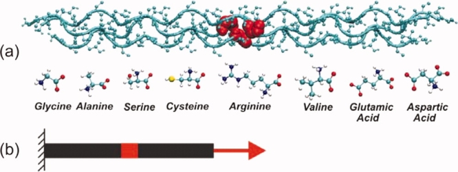
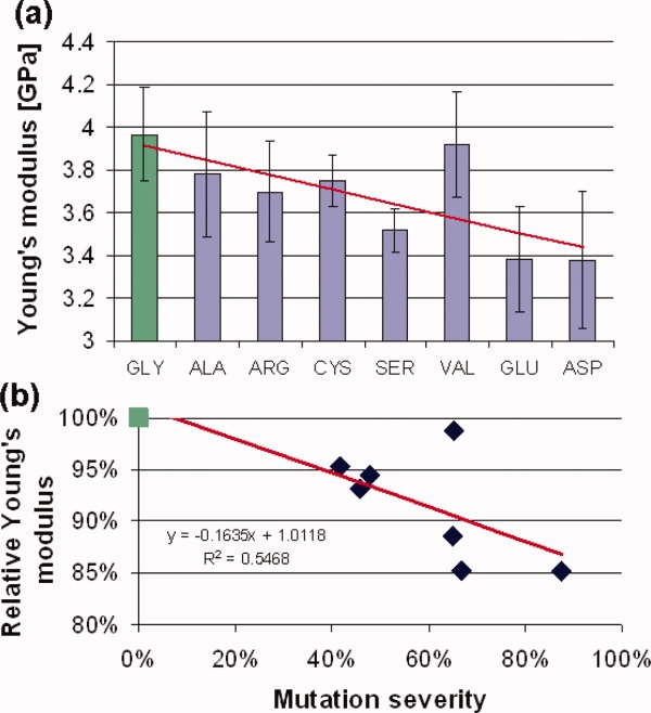
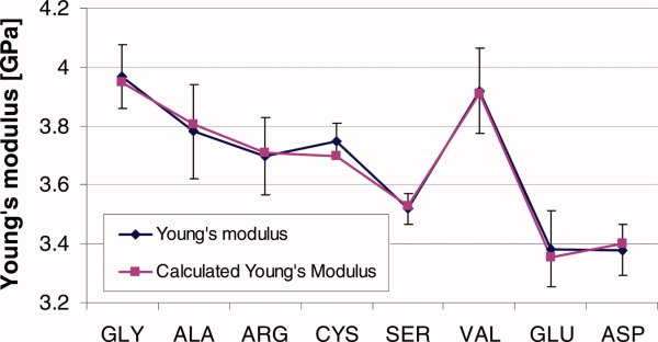
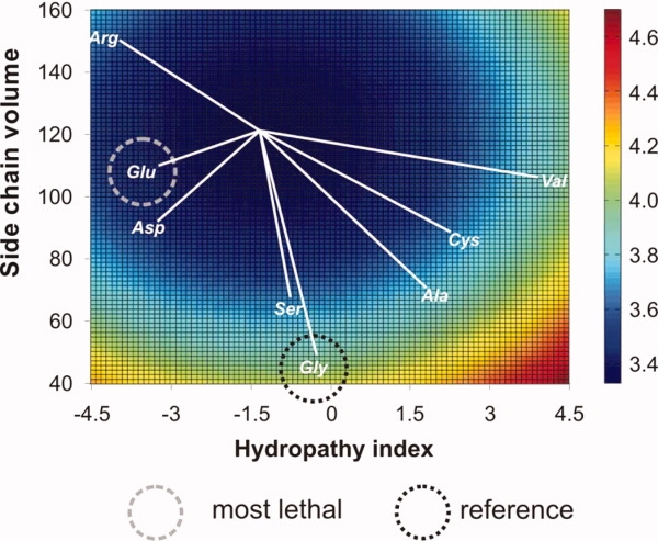
Osteogenesis imperfecta (OI) is a genetic disease characterized by fragile bones, skeletal deformities and, in severe cases, prenatal death that affects more than 1 in 10,000 individuals. Here we show by full atomistic simulation in explicit solvent that OI mutations have a significant influence on the mechanical properties of single tropocollagen molecules, and that the severity of different forms of OI is directly correlated with the reduction of the mechanical stiffness of individual tropocollagen molecules. The reduction of molecular stiffness provides insight into the molecular-scale mechanisms of the disease. The analysis of the molecular mechanisms reveals that physical parameters of side-chain volume and hydropathy index of the mutated residue control the loss of mechanical stiffness of individual tropocollagen molecules. We propose a model that enables us to predict the loss of stiffness based on these physical characteristics of mutations. This finding provides an atomistic-level mechanistic understanding of the role of OI mutations in defining the properties of the basic protein constituents, which could eventually lead to new strategies for diagnosis and treatment the disease. The focus on material properties and their role in genetic diseases is an important, yet so far only little explored, aspect in studying the mechanisms that lead to pathological conditions. The consideration of how material properties change in diseases could lead to a new paradigm that may expand beyond the focus on biochemical readings alone and include a characterization of material properties in diagnosis and treatment, an effort referred to as materiomics.
Figures
Similar articles
-
Molecular and mesoscale mechanisms of osteogenesis imperfecta disease in collagen fibrils.Biophys J. 2009 Aug 5;97(3):857-65. doi: 10.1016/j.bpj.2009.04.059. Biophys J. 2009. PMID: 19651044 Free PMC article.
-
Effect of changes in tropocollagen residue sequence and hydroxyapatite mineral texture on the strength of ideal nanoscale tropocollagen-hydroxyapatite biomaterials.J Mater Sci Mater Med. 2010 Jan;21(1):161-71. doi: 10.1007/s10856-009-3837-7. Epub 2009 Aug 5. J Mater Sci Mater Med. 2010. PMID: 19655234
-
Severity of osteogenesis imperfecta and structure of a collagen-like peptide modeling a lethal mutation site.Biochemistry. 2004 May 11;43(18):5314-23. doi: 10.1021/bi035676w. Biochemistry. 2004. PMID: 15122897
-
IFITM5 mutations and osteogenesis imperfecta.J Bone Miner Metab. 2016 Mar;34(2):123-31. doi: 10.1007/s00774-015-0667-1. Epub 2015 Jun 2. J Bone Miner Metab. 2016. PMID: 26031935 Review.
-
OIM and related animal models of osteogenesis imperfecta.Connect Tissue Res. 1995;31(4):265-8. doi: 10.3109/03008209509010820. Connect Tissue Res. 1995. PMID: 15612365 Review.
Cited by
-
A new paradigm for mechanobiological mechanisms in tumor metastasis.Semin Cancer Biol. 2012 Oct;22(5-6):385-95. doi: 10.1016/j.semcancer.2012.05.002. Epub 2012 May 18. Semin Cancer Biol. 2012. PMID: 22613484 Free PMC article. Review.
-
Variation in type I collagen fibril nanomorphology: the significance and origin.Bonekey Rep. 2013 Aug 21;2:394. doi: 10.1038/bonekey.2013.128. Bonekey Rep. 2013. PMID: 24422113 Free PMC article.
-
Nanoscale morphology of Type I collagen is altered in the Brtl mouse model of Osteogenesis Imperfecta.J Struct Biol. 2011 Jan;173(1):146-52. doi: 10.1016/j.jsb.2010.08.003. Epub 2010 Aug 7. J Struct Biol. 2011. PMID: 20696252 Free PMC article.
-
Nano-mechanical properties of individual mineralized collagen fibrils from bone tissue.J R Soc Interface. 2011 Apr 6;8(57):500-5. doi: 10.1098/rsif.2010.0413. Epub 2010 Oct 20. J R Soc Interface. 2011. PMID: 20961895 Free PMC article.
-
Collagen Gly missense mutations: Effect of residue identity on collagen structure and integrin binding.J Struct Biol. 2018 Sep;203(3):255-262. doi: 10.1016/j.jsb.2018.05.003. Epub 2018 May 11. J Struct Biol. 2018. PMID: 29758270 Free PMC article.
References
-
- Primorac D, Rowe DW, Mottes M, Barisic I, Anticevic D, Mirandola S, Lira MG, Kalajzic I, Kusec V, Glorieux FH. Osteogenesis imperfecta at the beginning of bone and joint decade. Croat Med J. 2001;42:393–415. - PubMed
-
- Rauch F, Glorieux FH. Osteogenesis imperfecta. Lancet. 2004;363:1377–1385. - PubMed
-
- Kozloff KM, Carden A, Bergwitz C, Forlino A, Uveges TE, Morris MD, Marini JC, Goldstein SA. Brittle IV mouse model for osteogenesis imperfecta IV demonstrates postpubertal adaptations to improve whole bone strength. J Bone Miner Res. 2004;19:614–622. - PubMed
Publication types
MeSH terms
Substances
LinkOut - more resources
Full Text Sources
Medical
