. 2009 Feb 24;48(7):1498-507.
doi: 10.1021/bi8020108.

A catalytic metal ion interacts with the cleavage Site G.U wobble in the HDV ribozyme

Affiliations

A catalytic metal ion interacts with the cleavage Site G.U wobble in the HDV ribozyme

Jui-Hui Chen et al. Biochemistry. .

Abstract

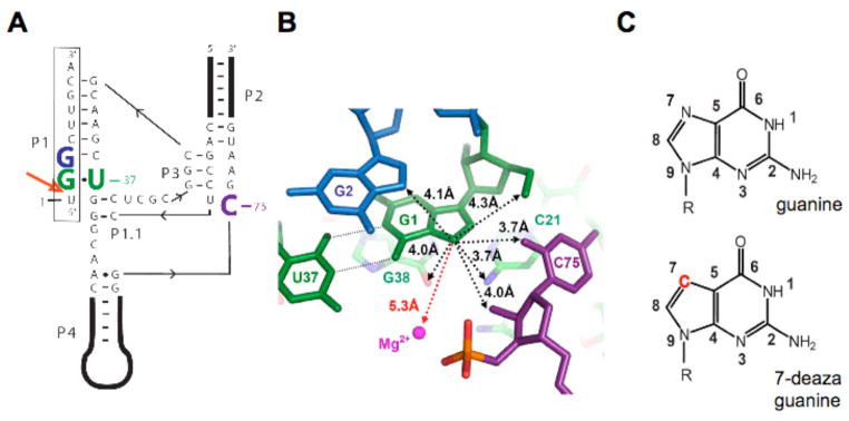
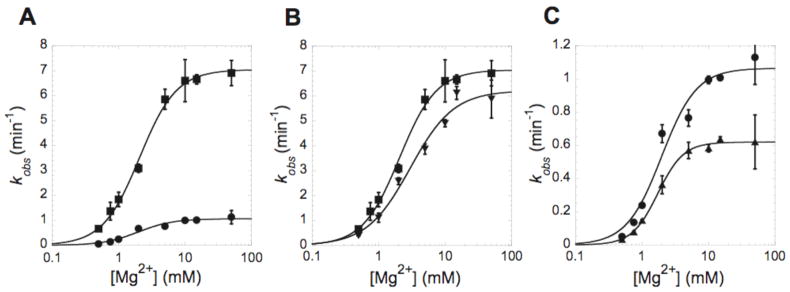
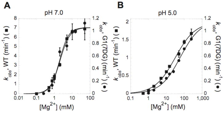
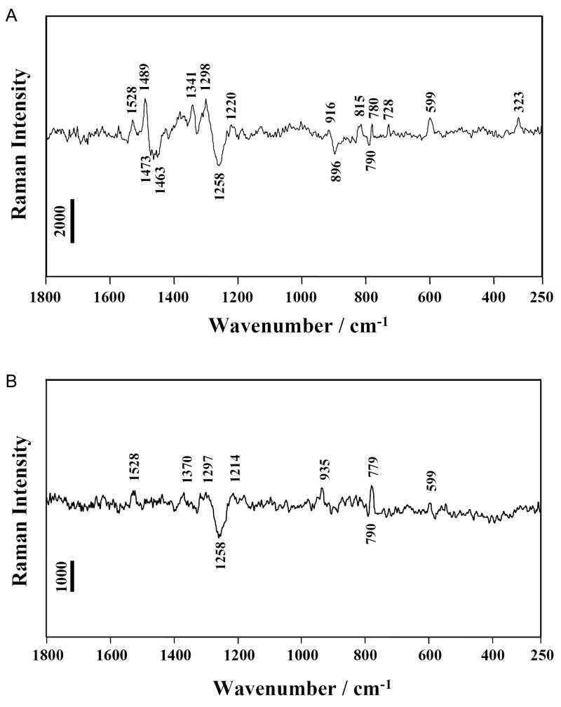
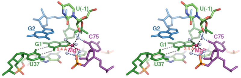
The HDV ribozyme self-cleaves by a chemical mechanism involving general acid-base catalysis to generate 2',3'-cyclic phosphate and 5'-hydroxyl termini. Biochemical studies from several laboratories have implicated C75 as the general acid and hydrated magnesium as the general base. We have previously shown that C75 has a pK(a) shifted >2 pH units toward neutrality [Gong, B., Chen, J. H., Chase, E., Chadalavada, D. M., Yajima, R., Golden, B. L., Bevilacqua, P. C., and Carey, P. R. (2007) J. Am. Chem. Soc. 129, 13335-13342], while in crystal structures, it is well-positioned for proton transfer. However, no evidence for a hydrated magnesium poised to serve as a general base in the reaction has been observed in high-resolution crystal structures of various reaction states and mutants. Herein, we use solution kinetic experiments and parallel Raman crystallographic studies to examine the effects of pH on the rate and Mg(2+) binding properties of wild-type and 7-deazaguanosine mutants of the HDV ribozyme. These data suggest that a previously unobserved hydrated magnesium ion interacts with N7 of the cleavage site G.U wobble base pair. Integrating this metal ion binding site with the available crystal structures provides a new three-dimensional model for the active site of the ribozyme that accommodates all available biochemical data and appears competent for catalysis. The position of this metal is consistent with a role of a magnesium-bound hydroxide as a general base as dictated by biochemical data.


Figures

Figure 1
Figure 1
Overview of the genomic HDV ribozyme structure. (A) Secondary structure of the genomic HDV ribozyme used in this study. The 9-nt substrate strand (boxed) is introduced in trans. The solid line with embedded arrow shows the 5′ to 3′ direction of chain connectivity in the ribozyme strand. The cleavage site between U(−1) and G1 is indicated with a heavy red arrow. Important nucleotides are colored as follows: C75 is purple, the cleavage site G•U wobble pair is green and G2 is blue. (B) Atoms surrounding the G1·U37 wobble pair in post-cleaved HDV ribozyme (PDB code1cx0)(5). Nucleotides are colored as in panel a. Non-hydrogen atoms within 4.5 Å to the N7 of G1 are shown by black dotted lines. The crystallographic metal ion found closest to G1 is 5.3 Å away and highlighted by a red dashed line. (c) The chemical structures of guanosine and 7-deazaguanosine. Modification in 7-deazaguanine relative to guanine is highlighted in red.
Figure 2
Figure 2
7-deazaguanosine at the cleavage site reduces ribozyme activity. Plots of kobs with varying concentrations of MgCl2. (A) Comparison of wild type (WT) (■) and G1(7DG) substrates (●). (B) WT (■) and G2(7DG) (▼) substrates. (C) G1(7DG) (●) and G1G2(7DG) substrates (▼). The cleavage reactions were performed at 37 °C in 25 mM Tris buffer pH 7.0. Each point is the average of three independent experiments and the standard deviation is shown as error bars. Data were fit to equation 2 described in methods. The resulting kinetic parameters are provided in Table 1.
Figure 3
Figure 3
Effect of 7-deazaguanosine on Mg2+ affinity is apparent at pH 5.0. Plots of kobs as a function of MgCl2 at (A) pH 7.0 and (B) pH 5.0. The cleavage reaction was performed at 37 °C in 25 mM Tris-Cl pH 7.0 or 25 mM sodium acetate pH 5.0. In each plot, the scale for kobs measured with the WT substrate (■) is shown on left Y axis, and the scale for kobs measured with the G1(7DG) (●) is shown on the right Y axis. Each point is the average of at least two independent experiments and the standard deviation is shown as error bars. Data were fit to equation 2. The resulting kinetic parameters are provided in Table 1.
Figure 4
Figure 4
Raman difference spectra. (A) [HDV pH 7.5] minus [HDV pH 5.0] (B) [HDV(G1(7DG)) pH 7.5] minus [HDV(G1(7DG)) pH 5.0]. The HDV ribozyme crystal was surrounded by cacodylate buffer pH 7.5 or acetate buffer pH 5.0 containing 20 mM Mg2+. Experimental details were described in previous Raman studies(17). Peak assignments are provided in Table S1. Vertical bars represent photon events.
Figure 5
Figure 5
The proposed model with hydrated metal ion close to the GU pair. Nucleotides are colored as in Figure 1A,B. C75 in purple, the cleavage site G•U wobble in green, and G2 in blue. The N7 of G1 interacts with a hydrated Mg2+ ion (magenta sphere) by inner-sphere coordination (shown as a red dashed line). The distance criteria used for inner-sphere (1.9–2.5Å) and outer-sphere (2.5–3.3Å) are based on the studies of Juneau and Cech(33). 2. Five water molecules (light blue sphere) surround the active site Mg2+ and complete the inner-sphere shell (1.9–2.1 Å, shown in black dash lines). The exact positions of the waters in this model are not known, however the water coordinates as drawn to provide a model of the expected radius of the hydration shell. The possible interaction between these coordinated waters and RNA functional groups are denoted by blue dotted lines. Other experimental criteria used to build the model are described in the text.

References

    1. Lai MM. The molecular biology of hepatitis delta virus. Annu Rev Biochem. 1995;64:259–286. - PubMed
    1. Been MD, Wickham GS. Self-cleaving ribozymes of hepatitis delta virus RNA. Eur J Biochem. 1997;247:741–753. - PubMed
    1. Wadkins TS, Been MD. Ribozyme activity in the genomic and antigenomic RNA strands of hepatitis delta virus. Cell Mol Life Sci. 2002;59:112–125. - PMC - PubMed
    1. Salehi-Ashtiani K, Luptak A, Litovchick A, Szostak JW. A genomewide search for ribozymes reveals an HDV-like sequence in the human CPEB3 gene. Science. 2006;313:1788–1792. - PubMed
    1. Ferre-D’Amare AR, Zhou K, Doudna JA. Crystal structure of a hepatitis delta virus ribozyme. Nature. 1998;395:567–574. - PubMed

Publication types