. 2009 Feb 17;106(7):2342-7.
doi: 10.1073/pnas.0813013106. Epub 2009 Jan 28.

The delta isoform of CaM kinase II is required for pathological cardiac hypertrophy and remodeling after pressure overload

Affiliations

The delta isoform of CaM kinase II is required for pathological cardiac hypertrophy and remodeling after pressure overload

Johannes Backs et al. Proc Natl Acad Sci U S A. .

Abstract

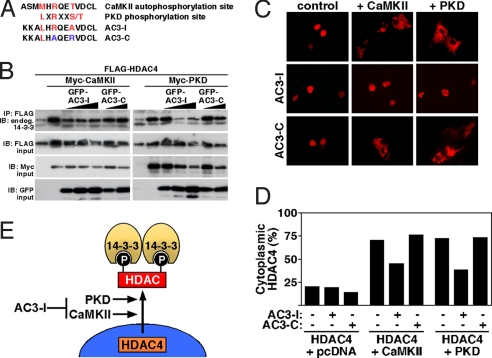
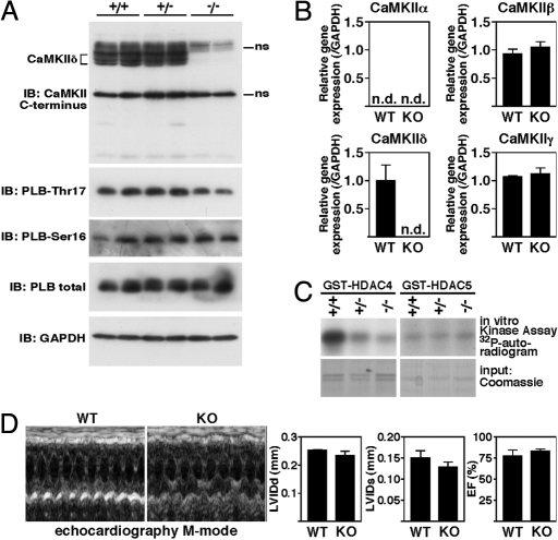
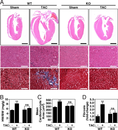
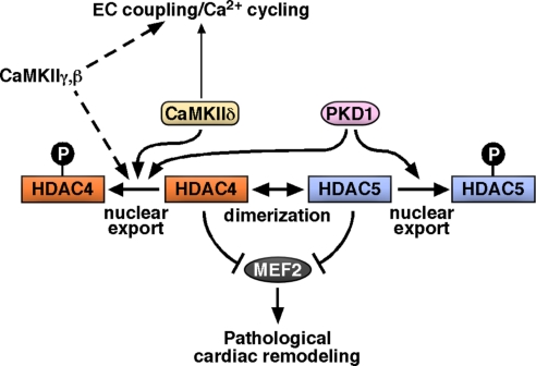
Acute and chronic injuries to the heart result in perturbation of intracellular calcium signaling, which leads to pathological cardiac hypertrophy and remodeling. Calcium/calmodulin-dependent protein kinase II (CaMKII) has been implicated in the transduction of calcium signals in the heart, but the specific isoforms of CaMKII that mediate pathological cardiac signaling have not been fully defined. To investigate the potential involvement in heart disease of CaMKIIdelta, the major CaMKII isoform expressed in the heart, we generated CaMKIIdelta-null mice. These mice are viable and display no overt abnormalities in cardiac structure or function in the absence of stress. However, pathological cardiac hypertrophy and remodeling are attenuated in response to pressure overload in these animals. Cardiac extracts from CaMKIIdelta-null mice showed diminished kinase activity toward histone deacetylase 4 (HDAC4), a substrate of stress-responsive protein kinases and suppressor of stress-dependent cardiac remodeling. In contrast, phosphorylation of the closely related HDAC5 was unaffected in hearts of CaMKIIdelta-null mice, underscoring the specificity of the CaMKIIdelta signaling pathway for HDAC4 phosphorylation. We conclude that CaMKIIdelta functions as an important transducer of stress stimuli involved in pathological cardiac remodeling in vivo, which is mediated, at least in part, by the phosphorylation of HDAC4. These findings point to CaMKIIdelta as a potential therapeutic target for the maintenance of cardiac function in the setting of pressure overload.


Conflict of interest statement

Conflict of interest statement: E.N.O. is cofounder of MiRagen Therapeutics. E.N.O. and J.B. are consultants for Gilead Therapeutics and have filed a patent on the modulation of cardiac hypertrophy and CaMKII.

Figures

Fig. 1.
Fig. 1.
AC3-I inhibits CaMKII and PKD. (A) Comparison of amino acid sequences of the CaMKII autophosphorylation site, the ideal PKD phosphorylation site, the so-called CaMKII inhibitory peptide (AC3-I), and its control peptide (AC3–3). (B and C). COS cells were transfected with FLAG-HDAC4, Myc-CaMKII, Myc-PKD1, and GFP-AC3-I or GFP-AC3-C as indicated. (B) Coimmunoprecipitation assays with COS cell lysates expressing the indicated proteins. Phosphorylation of HDAC4 was monitored by immunoprecipitation (IP) with anti-FLAG followed by immunoblotting (IB) with antibody against endogenous 14-3-3. Input proteins were detected by immunoblotting with antibodies against the indicated epitopes. (C) Representative immunocytochemistry showing the cellular localization of HDAC4 in cells transfected with FLAG-HDAC4, Myc-CaMKII, Myc-PKD1, and GFP-AC3-I or GFP-AC3-C as indicated. (D) Quantitative analysis of immunocytochemistry. (E) Illustration showing that HDAC4 is a common substrate of CaMKII and PKD, resulting in 14-3-3-mediated nuclear export. AC3-I inhibits both kinases.
Fig. 2.
Fig. 2.
Targeting of the mouse CaMKIIδ gene. (A) CaMKIIδ protein structure, intron–exon structure of the CaMKIIδ gene, and gene targeting strategy. LoxP sites were inserted in the introns flanking exons 1 and 2. Exon 2 encodes the ATP-binding motif required for kinase function. The neomycin resistance cassette (neo) was removed in the mouse germ line by breeding heterozygous mice to hACTB::FLPe transgenic mice, and deletion of exons 1 and 2 was achieved by breeding CaMKIIδloxP/loxP mice to CAG-Cre transgenic mice. (B) Representative Southern blot of genomic DNA from gene-targeted ES cells digested with PstI (P) using a probe hybridizing to a genomic region upstream (5′) of the long arm of the targeted region. The expected fragments were generated. (C) RT-PCR to detect WT and mutant CaMKIIδ (mut) transcripts, confirming that exons 1–4 are not transcribed in CaMKIIδ-KO mice. The reverse primer lies in exon 18, the numbers of the forward primers correspond to the exons containing their sequence.
Fig. 3.
Fig. 3.
Baseline characterization of CaMKIIδ-KO mice. (A) (Upper) Western blot analysis of endogenous CaMKII in cardiac extracts from CaMKIIδ+/+ (WT), CaMKIIδ+/− (HET), and CaMKIIδ−/− (KO) mice by using an antibody directed against the C terminus of all four CaMKII isoforms. (Lower) Western blot analysis of PLB at the CaMKII phosphorylation site Thr-17 and at the PKA phosphorylation site Ser-16 as well as total amounts of PLB and GAPDH as loading control; ns, nonspecific. (B) Transcripts for CaMKIIα, β, δ, and γ in hearts from WT and CaMKIIδ-KO mice were detected by quantitative PCR (n = 3 per group). Values indicate relative expression level to WT (±SEM). n.d., not detectable. (C) HDAC4 and HDAC5 kinase activity assays were performed in ventricular extracts from CaMKIIδ-KO mice and their WT littermates by using GST-HDAC4 and GST-HDAC5 substrates. HDAC4 but not HDAC5 phosphorylation was significantly decreased in ventricular extracts from CaMKIIδ-KO mice. Coomassie staining was used to demonstrate equivalent GST-HDAC4/HDAC5 input. (D) Transthoracic echocardiography revealed normal dimensions and function of CaMKIIδ-KO hearts. Shown are two representative M mode images and the quantitative analysis of the diastolic (LVIDd) and systolic (LVIDs) left ventricular internal diameter and ejection fraction (EF).
Fig. 4.
Fig. 4.
Diminished cardiac hypertrophy of CaMKIIδ-KO mice after TAC. CaMKIIδ-KO mice were subjected to either a sham operation (WT and KO mice, n = 4) or TAC (WT and KO, n = 6). Hearts for analysis were taken 21 days after sham or TAC. (A) Histological sections stained with H&E and Masson's trichrome to detect fibrosis. [Scale bars, 2 mm (Upper) and 100 μm (Lower).] (B) HW/BW ratios (±SEM) of WT and CaMKIIδ-KO mice. (C) Mean cross-sectional area of cardiomyocytes (±SEM). (D) Quantification of fibrosis. Values indicate fold changes of fibrosis in each group compared with a group of sham-operated WT mice (±SEM); ns (nonsignificant), P > 0.05; **, P < 0.001.
Fig. 5.
Fig. 5.
Single-cell experiments. (A) Typical cells from WT and CaMKIIδ-KO mice under basal conditions and 6 weeks after TAC. (B) Under basal conditions, cell volume is not different between WT and KO mice. After TAC, both WT and KO myocytes show cellular hypertrophy. However, this hypertrophy is significantly diminished in KO-TAC compared with WT-TAC. (C) Representative Ca2+ transients from WT and KO animals both under basal conditions and after TAC. (D) Ca2+ transient amplitude (ΔF/F0) is slightly increased in KO myocytes under basal conditions but is not different between WT and KO after TAC. (E) SR Ca2+ load as measured by rapid application of caffeine is not different between KO and WT under basal conditions or after TAC; n.s. (nonsignificant), P > 0.05; *, P < 0.05.
Fig. 6.
Fig. 6.
Activation of fetal genes in CaMKIIδ-KO mice after TAC. Transcripts for markers of hypertrophy in hearts from WT and CaMKIIδ-KO mice were detected by quantitative PCR 21 days after TAC (n = 3 per group). Values indicate relative expression level to a WT sham-operated group (±SEM). ANP, atrial natriuretic peptide; BNP, brain natriuretic peptide; Myh7, βMHC, myosin heavy chain; n.s. (nonsignificant), P > 0.05; *, P < 0.05; **, P < 0.001.
Fig. 7.
Fig. 7.
Model of molecular mechanism. CaMKIIδ is required for pathological cardiac remodeling at least in part by phosphorylating HDAC4. Regarding EC coupling, redundant roles of other CaMKII isoforms expressed in the heart need to be evaluated. PKD1 also phosphorylates class IIa HDACs and induces pathological cardiac remodeling. Target-specific inhibition (e.g., disrupting the CaMKII–HDAC4 interaction) might be a way to inhibit the CaMKII effects on pathological cardiac remodeling without affecting possible essential CaMKII functions.

References

    1. Saito Y, et al. Augmented expression of atrial natriuretic polypeptide gene in ventricle of human failing heart. J Clin Invest. 1989;83:298–305. - PMC - PubMed
    1. Oka T, Xu J, Molkentin JD. Reemployment of developmental transcription factors in adult heart disease. Semin Cell Dev Biol. 2007;18:117–131. - PMC - PubMed
    1. Frey N, Olson EN. Cardiac hypertrophy: The good, the bad, and the ugly. Annu Rev Physiol. 2003;65:45–79. - PubMed
    1. Frey N, McKinsey TA, Olson EN. Decoding calcium signals involved in cardiac growth and function. Nat Med. 2000;6:1221–1227. - PubMed
    1. McKinsey TA. Derepression of pathological cardiac genes by members of the CaM kinase superfamily. Cardiovasc Res. 2007;73:667–677. - PubMed

Publication types

MeSH terms