. 2009 Apr 16;113(16):3875-84.
doi: 10.1182/blood-2008-09-177055. Epub 2009 Jan 28.

Breaking tolerance to self, circulating natural killer cells expressing inhibitory KIR for non-self HLA exhibit effector function after T cell-depleted allogeneic hematopoietic cell transplantation

Affiliations

Breaking tolerance to self, circulating natural killer cells expressing inhibitory KIR for non-self HLA exhibit effector function after T cell-depleted allogeneic hematopoietic cell transplantation

Junli Yu et al. Blood. .

Abstract

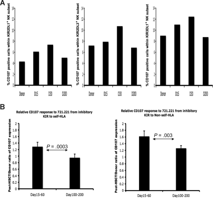
Alloreactive natural killer (NK) cells are an important influence on hematopoietic stem cell transplantation (HSCT) outcome. In HLA-mismatched HSCT, alloreactivity occurs when licensed donor NK cells expressing inhibitory killer Ig-like receptors (KIR) for donor MHC class I ligands recognize the lack of the class I ligands in the mismatched recipient ("missing self"). Studies in HLA-matched HSCT, however, have also demonstrated improved outcome in patients lacking class I ligands for donor inhibitory KIR ("missing ligand"), indicating that classically nonlicensed donor NK cells expressing KIR for non-self MHC class I ligands may exhibit functional competence in HSCT. We examined NK function in 16 recipients of T cell-depleted allografts from HLA-identical or KIR-ligand matched donors after myeloablative therapy. After HSCT, nonlicensed NK cells expressing inhibitory KIR for non-self class I exhibit robust intracellular IFN-gamma and cytotoxic response to target cells lacking cognate ligand, gradually becoming tolerized to self by day 100. These findings could not be correlated with cytokine environment or phenotypic markers of NK development, nor could they be attributed to non-KIR receptors such as CD94/NKG2A. These findings confirm that NK alloreactivity can occur in HLA-matched HSCT, where tolerance to self is either acquired by the stem cell-derived NK cell after exiting the bone marrow or where tolerance to self can be temporarily overcome.


Figures

Figure 1
Figure 1
Function of NK cells expressing inhibitory KIR is altered immediately after TCD-HSCT. Shown is the percentage of IFN-γ–producing cells among KIR-expressing NK populations from the HLA-C1/C1, Bw6/Bw6 donor, the HLA-identical patient (patient no. 2) before transplantation, and the patient at day +30, day +60, and day +200 after HSCT after coincubation with the indicated target cells. (A) NK cells exclusively expressing the inhibitory KIR for self-HLA class I (S-KIR) KIR2DL3 after HSCT are hyperresponsive compared with the donor and patient pre-HSCT, normalizing to donor level by day +200, and reactivity is inhibited upon challenge with the cognate class I ligand HLA-Cw3. NK cells exclusively expressing the inhibitory KIR for the non-self HLA class I (NS-KIR) KIR2DL1 (B) or KIR3DL1 (C) are functionally competent compared with the donor and patient pre-HSCT, reacting to lack of ligand. Reactivity is inhibited upon challenge with the cognate class I ligands HLA-Cw4 or Bw4, respectively.
Figure 2
Figure 2
Aggregate NK function from 16 TCD-HSCT donor-recipient pairs. IFN-γ response to class I–negative 721.221 target cells was calculated relative to baseline for each inhibitory KIR in each of 16 TCD-HSCT donor-recipient pairs by the formula (KIR+IFN-γ+/KIR+)/(KIRIFN-γ+/KIR). The relative response of NK cells exclusively expressing specific inhibitory KIR was compared among the allograft donor, the patient at day +15 to 60, and the patient at days +100 to +200 after HSCT. (A) NK cells exclusively expressing inhibitory KIR to self-HLA from patients at days +15 to +60 are hyperresponsive compared with their donors (P = .004), normalizing to donor levels after day +100. (B) NK cells exclusively expressing inhibitory KIR to non-self HLA from patients at days +15 to +60 after HSCT are functionally competent (P < .001) compared with the donor, decreasing but still higher than donor by days +100 to +200 (P < .001). The paired Student t test was performed to compute the P value for these comparisons. NK cells expressing KIR for non-self HLA from days +15 to +60 NK cells are comparable in effector response to steady-state healthy donor NK cells expressing KIR for self-HLA (*).
Figure 3
Figure 3
Nonlicensed NK cells expressing inhibitory KIR for non-self class I after TCD-HSCT are activated in response to leukemic cell lines and primary leukemic blasts lacking cognate ligands. Percentages of IFN-γ–producing cells among nonlicensed NK cells exclusively expressing the inhibitory KIR for non-self HLA class I ligand (NS-KIR) after activation by leukemic targets are shown for 2 patients. Targets include the patient-derived ALL cell line PhALL3.1 (HLA-C1/C1; Bw6/Bw6), the AML cell lines HL60 (HLA-C2/C2; Bw4/Bw6) and KASUMI-1 (HLA-C1/C1; Bw4/Bw6), and primary biphenotypic leukemic blasts PBL-1 (HLA-C1/C1; Bw4/Bw4). (A) Compared with the donor control, NK cells exclusively expressing NS-KIR KIR2DL3 from the HLA-C2/C2, Bw4/Bw4 post-HSCT patient (patient no. 5) are specifically activated against HL60, which lacks the cognate HLA-C1 ligand. (B) NK cells exclusively expressing NS-KIR KIR2DL1 from the HLA-C1/C1, Bw4/Bw4 post-HSCT recipient (patient no. 16) are activated against PhALL3.1, KASUMI-1, and PBL-1, which lack the cognate HLA-C2 ligand.
Figure 4
Figure 4
Licensed and nonlicensed NK-cell degranulation after HSCT. Donor and post-HSCT patient PBMCs (patients 1, 8, 9) were incubated with 721.221 target cells and measured for CD107a expression. Degranulation was assessed as the percentage of CD107a+ cells within NK populations expressing specific KIR. (A) Percentage of CD107a-positive cells within the NK cells exclusively expressing NS-KIR KIR2DL1 (left), S-KIR KIR2DL3 (middle), and S-KIR KIR3DL1 (right) from the healthy HLA-C1/C1, Bw4/Bw6 HSCT donor (no. 9), the HLA-C1/C1, Bw4/Bw6 patient at day +15, day +30, and day +200 after HSCT. Findings are representative of studies performed in 3 donor-recipient pairs. (B) The post-HSCT/pre-HSCT ratio of CD107a expression was compared in the patient at days +15 to +60 and days +100 to +200 after HSCT (n = 3). The paired Student t test was performed to compute the P value for these comparisons.
Figure 5
Figure 5
Unlicensed KIR+NKG2A NK cells after HSCT demonstrate functional competence. IFN-γ response to class I–negative 721.221 target cells was calculated relative to baseline for each inhibitory KIR in each of 3 TCD-HSCT donor-recipient pairs (nos. 10, 12, and 14) by the formula (NKG2AKIR+IFN-γ+/NKG2AKIR+)/(NKG2AKIRIFN-γ+/NKG2AKIR). The relative response of NK cells exclusively expressing specific inhibitory KIR was compared between the allograft donor and the patient at days +100 to +200 after HSCT. (A) CD94/NKG2A-negative NK cells exclusively expressing inhibitory KIR to self-HLA from patients at days +100 to +200 are slightly hyporesponsive compared with their donors (P = .02). (B) CD94/NKG2A-negative NK cells exclusively expressing inhibitory KIR to non-self HLA from patients demonstrate higher response compared with the donor (P < .01). The paired Student t test was performed to compute the P value for these comparisons.
Figure 6
Figure 6
Majority of post-HSCT KIR+ NK cells belong to the CD56dim NK subset. PBMC preparations from the patient at day +15 after HSCT and day +100 after HSCT were stained with monoclonal antibodies specific for CD3, CD56, KIR2DL1, KIR2DL3, KIR3DL1, CD16, and CD117. (A) Flow cytometric staining gated on CD3CD56+ NK cells demonstrates that the overwhelming majority of KIR+ cells are CD56dim at both time points. Data are representative of analysis from 15 patients. (B) Flow cytometric staining gated on CD3CD56+ KIR+ cells shows the majority of cells at both time points are CD117CD16+. Data are representative of analysis from 6 patients.

References

    1. Biron CA, Nguyen KB, Pien GC, Cousens LP, Salazar-Mather TP. Natural killer cells in antiviral defense: function and regulation by innate cytokines. Annu Rev Immunol. 1999;17:189–220. - PubMed
    1. Trinchieri G. Biology of natural killer cells. Adv Immunol. 1989;47:187–376. - PMC - PubMed
    1. Ljunggren HG, Karre K. In search of the missing self: MHC molecules and NK cell recognition. Immunol Today. 1990;11:237–244. - PubMed
    1. Colonna M, Samaridis J. Cloning of immunoglobulin-superfamily members associated with HLA-C and HLA-B recognition by human natural killer cells. Science. 1995;268:405–408. - PubMed
    1. Long EO. Regulation of immune responses through inhibitory receptors. Annu Rev Immunol. 1999;17:875–904. - PubMed

Publication types

MeSH terms