. 2009 Feb 17;106(7):2331-6.
doi: 10.1073/pnas.0805159106. Epub 2009 Jan 29.

R-Spondin1 protects mice from chemotherapy or radiation-induced oral mucositis through the canonical Wnt/beta-catenin pathway

Affiliations

R-Spondin1 protects mice from chemotherapy or radiation-induced oral mucositis through the canonical Wnt/beta-catenin pathway

Jingsong Zhao et al. Proc Natl Acad Sci U S A. .

Abstract

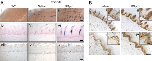
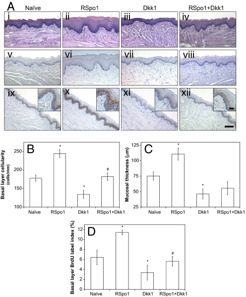
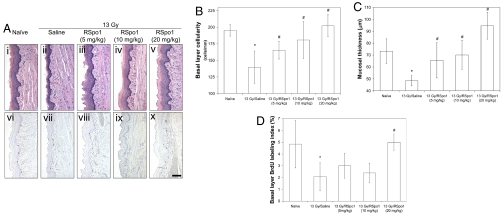
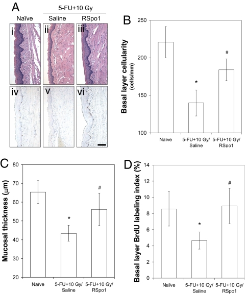
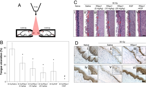
R-Spondin1 (RSpo1) is a novel secreted protein that augments canonical Wnt/beta-catenin signaling. We injected recombinant RSpo1 protein into transgenic Wnt reporter TOPGAL mice and have identified the oral mucosa as a target tissue for RSpo1. Administration of RSpo1 into normal mice triggered nuclear translocation of beta-catenin and resulted in increased basal layer cellularity, thickened mucosa, and elevated epithelial cell proliferation in tongue. We herein evaluated the therapeutic potential of RSpo1 in treating chemotherapy or radiotherapy-induced oral mucositis in several mouse models. Prophylactic treatment with RSpo1 dose-dependently overcame the reduction of basal layer epithelial cellularity, mucosal thickness, and epithelial cell proliferation in tongues of mice exposed to whole-body irradiation. RSpo1 administration also substantially alleviated tongue mucositis in the oral cavity of mice receiving concomitant 5-fluorouracil and x-ray radiation. Furthermore, RSpo1 significantly reduced the extent of tongue ulceration in mice receiving a single fraction, high dose head-only radiation in a dose-dependent manner. Moreover, combined therapy of RSpo1 and keratinocyte growth factor resulted in complete healing of tongue ulcers in mice subjected to snout-only irradiation. In conclusion, our results demonstrate RSpo1 to be a potent therapeutic agent for oral mucositis by enhancing basal layer epithelial regeneration and accelerating mucosal repair through up-regulation of Wnt/beta-catenin pathway.


Conflict of interest statement

The authors declare no conflict of interest.

Figures

Fig. 1.
Fig. 1.
Effect of RSpo1 on tongue mucosa in TOPGAL mice. (A) (i–iii) Whole-mount Bluo-Gal-stained mouse dorsal tongues. (Scale bar, 0.5 mm.) (iv–vi) Coronal sections of Bluo-Gal-stained mouse dorsal tongues with nuclear fast red counterstain. (Scale bar, 100 μm.) (vii–ix) BrdU immunocytochemistry of mouse dorsal tongue mucosa. (Scale bar, 100 μm.) (i, iv, and vii) WT, (ii, v, and viii) TOPGAL treated with saline, (iii, vi, and ix) TOPGAL treated with RSpo1. (B) β-catenin immunohistochemical study in dorsal (i and ii) and ventral (iii and iv) tongues of TOPGAL mice. (i and iii) Saline; (ii and iv) RSpo1. (Scale bar, 100 μm; Insets, 25 μm.)
Fig. 2.
Fig. 2.
Effect of RSpo1 and/or Dkk1 on mouse tongue mucosa. (A) H&E staining (i–iv) BrdU immunohistochemistry, (v–viii) and β-catenin immunohistochemistry (ix–xii) of mouse ventral tongue mucosa. i, v, and ix, naïve; ii, vi, and x, RSpo1; iii, vii, and xi, Dkk1; viii, and iv, RSpo1+Dkk1. (Scale bar, 100 μm; Insets, 25 μm .) (B) Basal layer epithelial cellularity in mouse ventral tongue mucosa. (C) Mucosal thickness in mouse ventral tongue. (D) Basal epithelial layer BrdU labeling index in mouse ventral tongue. *P < 0.05 (RSpo1 vs. naïve, Dkk1 vs. naïve); #P < 0.05 (RSpo1+Dkk1 vs. Dkk1).
Fig. 3.
Fig. 3.
Effect of RSpo1 on tongue mucosa in whole-body irradiated mice. (A) Microscopic images of H&E staining (i–v) and BrdU immunohistochemistry (vi–x) of ventral tongue mucosa in mice. i and vi, naïve; ii and vii, 13 Gy/saline; iii and viii, 13 Gy/RSpo1 (5 mg/kg); iv and ix, 13 Gy/RSpo1 (10 mg/kg); v and x, 13 Gy/RSpo1 (20 mg/kg). (Scale bar, 100 μm.) (B) Basal layer epithelial cellularity in ventral tongue mucosa in mice. (C) Mucosal thickness in ventral tongue in mice. (D) BrdU labeling index in basal epithelial layer of mouse ventral tongue. *P < 0.05 (13 Gy/saline vs. naïve); #P < 0.05 (13 Gy/RSpo1 vs. 13 Gy/saline).
Fig. 4.
Fig. 4.
Characterization of RSpo1 on tongue mucosa in mice receiving combined treatment of 5-FU and whole-body radiation. (A) Representative microscopic images of H&E staining (i–iii) and BrdU immunohistochemistry (iv–vi) from mouse ventral tongue mucosa. (i and iv) Naïve, (ii and v) (5-FU + 10 Gy)/saline, (iii and vi) (5-FU + 10 Gy)/RSpo1. (Scale bar, 100 μm.) (B) Epithelial cellularity of ventral tongue mucosal basal layer in mice. (C) Ventral tongue mucosal thickness in mice. (D) Epithelial cell BrdU labeling index in mouse ventral tongue mucosal basal layer. *P < 0.05 [(5-FU + 10 Gy)/saline vs. naïve]; #P < 0.05 [(5-FU + 10 Gy)/RSpo1 vs. (5-FU + 10 Gy)/saline].
Fig. 5.
Fig. 5.
Activity of RSpo1 on tongue ulceration in mice treated with high dose head-only irradiation. (A) Schematic illustration of setup of head-only radiation for mice. Red triangle represents passage of x-ray irradiation. (B) Overt ulceration in mouse tongue mucosa. *P < 0.05 (30 Gy/RSpo1 or 30 Gy/KGF vs. 30 Gy/saline); #P < 0.05 [30 Gy/(RSpo1+KGF) vs. 30 Gy/saline, 30 Gy/RSpo1, or 30 Gy/KGF]. (C) Histological representations of mouse dorsal tongue (H&E staining). Red lines donate tongue ulceration with complete mucosal denudation. (i) Naïve, (ii) 30 Gy/saline, (iii) 30 Gy/RSpo1 (5 mg/kg), (iv) 30 Gy/RSpo1 (10 mg/kg), (v) 30 Gy/RSpo1 (20 mg/kg), (vi) 30 Gy/KGF, (vii) 30 Gy/(RSpo1+KGF). (Scale bar, 100 μm.) (D) β-catenin immunohistochemical staining in mouse dorsal (i–iii) and ventral (v and vi) tongues. (i and iv) Naïve, (ii and v) 30 Gy/saline, (iii and vi) 30 Gy/RSpo1. (Scale bar, 100 μm; Insets, 25 μm.)

References

    1. Kim KA, et al. R-Spondin proteins: A novel link to beta-catenin activation. Cell Cycle. 2006;5:23–26. - PubMed
    1. Nam JS, Turcotte TJ, Smith PF, Choi S, Yoon JK. Mouse cristin/R-spondin family proteins are novel ligands for the Frizzled 8 and LRP6 receptors and activate beta-catenin-dependent gene expression. J Biol Chem. 2006;281:13247–13257. - PubMed
    1. Wei Q, et al. R-spondin1 is a high affinity ligand for LRP6 and induces LRP6 phosphorylation and beta-catenin signaling. J Biol Chem. 2007;282:15903–15911. - PubMed
    1. Binnerts ME, et al. R-Spondin1 regulates Wnt signaling by inhibiting internalization of LRP6. Proc Natl Acad Sci USA. 2007;104:14700–14705. - PMC - PubMed
    1. Kamata T, et al. R-spondin, a novel gene with thrombospondin type 1 domain, was expressed in the dorsal neural tube and affected in Wnts mutants. Biochim Biophys Acta. 2004;1676:51–62. - PubMed

LinkOut - more resources