. 2009 Apr;77(4):1337-48.
doi: 10.1128/IAI.01456-08. Epub 2009 Jan 29.

Helicobacter pylori-induced interleukin-12 p40 expression

Affiliations

Helicobacter pylori-induced interleukin-12 p40 expression

Eriko Takeshima et al. Infect Immun. 2009 Apr.

Retraction in

  • Retraction. Helicobacter pylori-induced interleukin-12 p40 expression.
    Takeshima E, Tomimori K, Teruya H, Ishikawa C, Senba M, D'Ambrosio D, Kinjo F, Mimuro H, Sasakawa C, Hirayama T, Fujita J, Mori N. Takeshima E, et al. Infect Immun. 2011 Jan;79(1):546. doi: 10.1128/IAI.01069-10. Infect Immun. 2011. PMID: 21177935 Free PMC article. No abstract available.

Abstract

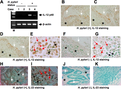
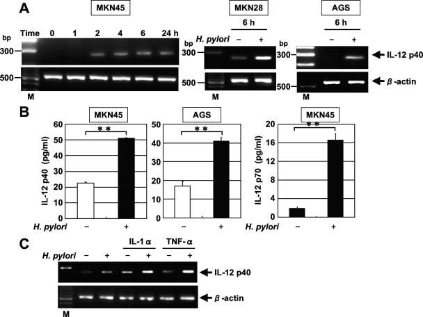
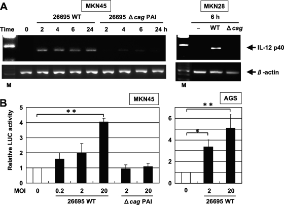
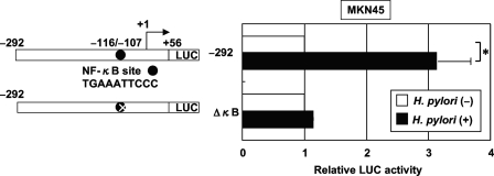
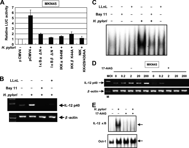
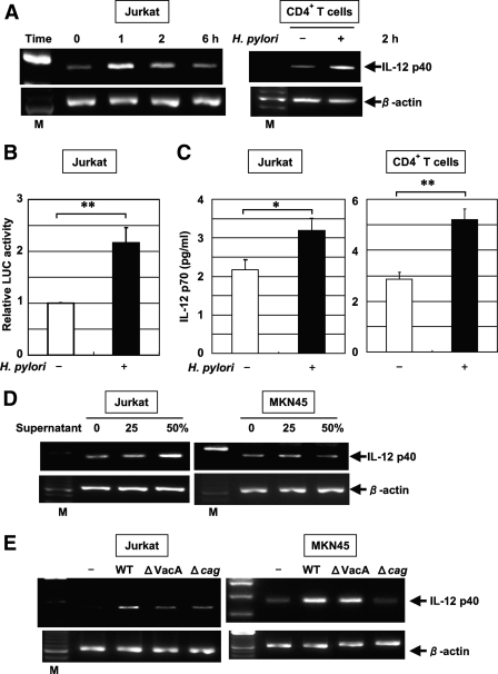
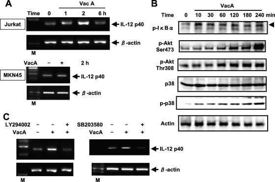
Interleukin-12 (IL-12) is a heterodimeric cytokine produced by antigen-presenting cells that promotes the development of T-helper lymphocyte 1 (Th1). Chronic gastritis induced by Helicobacter pylori is considered a Th1-mediated process. IL-12 levels in gastric biopsy samples of H. pylori-infected patients are higher than in those of uninfected individuals, but the cellular source of IL-12 remains elusive. IL-12 staining was detected in mucosal epithelial cells, lymphocytes, and macrophages in specimens of patients with H. pylori-positive gastritis. Therefore, we investigated IL-12 p40 mRNA induction by H. pylori in gastric epithelial cells and T cells. Although cag pathogenicity island (PAI)-positive H. pylori induced IL-12 p40 mRNA expression, an isogenic mutant of the cag PAI failed to induce it in both cell types. Supernatants from H. pylori cultures and H. pylori VacA induced IL-12 p40 mRNA expression in T cells but not in epithelial cells. The activation of the IL-12 p40 promoter by H. pylori was mediated through NF-kappaB. The transfection of IkappaB kinase and NF-kappaB-inducing kinase dominant-negative mutants inhibited H. pylori-induced IL-12 p40 activation. Inhibitors of NF-kappaB, phosphatidylinositol 3-kinase, p38 mitogen-activated protein kinase, and Hsp90 suppressed H. pylori- and VacA-induced IL-12 p40 mRNA expression. The results indicate that H. pylori induces IL-12 p40 expression by the activation of NF-kappaB, phosphatidylinositol 3-kinase, and p38 mitogen-activated protein kinase. Hsp90 is also a crucial regulator of H. pylori-induced IL-12 p40 expression. In addition to the cag PAI, VacA might be relevant in the induction of IL-12 expression and a Th1-polarized response only in T cells.


Figures

FIG. 1.
FIG. 1.
Expression of IL-12 p40 in H. pylori-infected gastric mucosa. (A) RT-PCR analysis of IL-12 p40 in representative human gastric tissues. Lanes 1 and 2, normal mucosa; lanes 3 and 4, H. pylori-positive gastritis; lane M, markers. β-Actin expression served as a control. Immunohistochemical detection of IL-12 (B to G) and IL-23 (H to K) in tissues of patients with H. pylori-positive gastritis. Serial sections of gastric biopsy specimens were stained with mouse monoclonal antibodies to IL-12 and IL-23 and counterstained with methyl green. Also shown are representative examples of mucosa from patients with H. pylori-positive gastritis (B to E, H, and I) and normal mucosa (F, G, J, and K). Note the positive staining for IL-12 and IL-23 in epithelial cells and lymphocytes as well as macrophages from patients with H. pylori-positive gastritis. (B to D and F) Original magnification, ×170. (E and G) Original magnification, ×430. (H, J, and K) Original magnification, ×140. (I) Original magnification, ×360. The white, black, and red arrows indicate the surfaces of epithelial cells, lymphocytes, and macrophages, respectively. The asterisks indicate deeper structures of epithelial cells.
FIG. 2.
FIG. 2.
H. pylori-induced IL-12 p40 mRNA expression and secretion in gastric epithelial cells. (A) Dynamics of H. pylori-induced IL-12 p40 mRNA expression. Total RNA was extracted from the indicated cells that had been infected with H. pylori ATCC 49503 for the indicated times and used for RT-PCR (MOI, 20). Representative results of three similar experiments in each panel are shown. M, size marker. (B) Increased secretion of IL-12 p40 and p70 into the supernatants of MKN45 and AGS cell cultures in response to H. pylori ATCC49503 infection at 24 h. IL-12 p40 and p70 concentrations in the supernatants were determined by ELISA. Data are means ± standard deviations from three experiments. **, P < 0.01, as determined by the Student t test. (C) Expression of IL-12 p40 mRNA was further upregulated in response to H. pylori when there was stimulation by the proinflammatory cytokines IL-1α and TNF-α in MKN45 cells. MKN45 cells were stimulated with IL-1α (100 ng/ml) or TNF-α (100 ng/ml) for 12 h and then incubated in the presence or absence of H. pylori for 2 h, and the expression of IL-12 p40 mRNA was assessed by RT-PCR. Representative results from three similar experiments are shown in each panel.
FIG. 3.
FIG. 3.
cag PAI products of H. pylori are required for the induction of IL-12 p40 mRNA expression. (A) Total RNA was extracted from the indicated cells that had been infected with the wild-type strain 26695 (WT) or the isogenic mutant Δcag PAI (Δcag) for the indicated times and used for RT-PCR. Representative results of three similar experiments are shown in each panel. M, size marker. (B) H. pylori infection increased IL-12 p40 promoter activity in a dose-dependent fashion. A luciferase (LUC) reporter construct was transfected into MKN45 and AGS cells, and the cells subsequently were infected with the WT or Δcag PAI for 6 h. The activities are expressed relative to that of cells transfected with the reporter construct without further H. pylori infection, which was defined as 1. Data are means ± standard deviations from three independent experiments. *, P < 0.05; **, P < 0.01; both were determined by the Student t test.
FIG. 4.
FIG. 4.
H. pylori activates the IL-12 p40 promoter through the NF-κB binding site. (Left) Schematic diagram of the IL-12 p40 reporter constructs containing the wild type (−292) and internal deletion mutant of the NF-κB site (ΔκB). LUC, luciferase. (Right) Either the IL-12 p40 reporter construct or a construct bearing an internal deletion of the NF-κB binding site was transfected into MKN45 cells, and subsequently the cells were infected with H. pylori ATCC 49503 for 6 h (MOI, 20:1). The activity is expressed relative to that of cells transfected with each construct without further H. pylori infection, which was defined as 1. Data are means ± standard deviations from three independent experiments. *, P < 0.05, as determined by the Student t test.
FIG. 5.
FIG. 5.
H. pylori infection induces NF-κB binding activity. (A) Time course of NF-κB activation in MKN45, MKN28, and AGS cells infected with H. pylori, as evaluated by EMSA (left). Nuclear extracts from the indicated cells infected with H. pylori ATCC 49503 for the indicated times were mixed with 32P-labeled NF-κB probe (MOI, 20:1). Competition assays were performed with nuclear extracts from these cells infected with H. pylori ATCC 49503 for 30 min (right). Where indicated, 100-fold excess amounts of each specific competitor oligonucleotide were added to the reaction mixture with labeled probe NF-κB (lanes 2 to 4). A supershift assay of NF-κB DNA binding complexes in the same nuclear extracts also was performed. Where indicated, appropriate antibodies (Ab) were added to the reaction mixture before the addition of the 32P-labeled probe (lanes 5 to 9). Arrows indicate the specific complexes, while arrowheads indicate the DNA binding complexes supershifted by antibodies. (B) cag PAI products of H. pylori are required for the induction of NF-κB binding activity in MKN45 cells. Nuclear extracts from MKN45 cells infected with different densities (MOI) of wild-type strain 26695 or the isogenic mutant Δcag PAI for 1 h were analyzed for NF-κB. Representative results of three similar experiments are shown in each panel. WT, wild type.
FIG. 6.
FIG. 6.
NF-κB signal is essential for the activation of IL-12 p40 expression by H. pylori in gastric epithelial cells. (A) Functional effects of IκBα and IκBβ dominant-interfering mutants and kinase-deficient IKKα, IKKβ, and NIK mutants on the H. pylori-induced activation of the IL-12 p40 promoter. MKN45 cells were transfected with the IL-12 p40 reporter construct and the indicated mutant plasmids or empty vector (pCMV4) and then infected with H. pylori ATCC 49503 for 6 h. The open bar indicates the luciferase (LUC) activity of the IL-12 p40 reporter construct and pCMV4 without H. pylori infection. All values first were calculated as the change (n-fold) in induction values relative to the basal level measured in uninfected cells. Data are means ± standard deviations from three independent experiments. (B) Bay 11-7082 and LLnL inhibit IL-12 p40 mRNA expression induced by H. pylori. MKN45 cells were pretreated with Bay 11-7082 (20 μM) or LLnL (20 μM) for 1 h prior to H. pylori infection and subsequently were infected with H. pylori ATCC 49503 for 6 h. IL-12 p40 mRNA expression on harvested cells was analyzed by RT-PCR. (C) Bay 11-7082 and LLnL inhibit H. pylori-induced NF-κB DNA binding. MKN45 cells were pretreated with Bay 11-7082 (20 μM) or LLnL (20 μM) for 1 h prior to H. pylori infection and subsequently were infected with H. pylori ATCC 49503 for 1 h. Nuclear extracts from harvested cells were analyzed for NF-κB. (D) Inhibitory effects of 17-AAG on H. pylori-induced IL-12 p40 expression. MKN45 cells were incubated with 1 μM 17-AAG for 16 h prior to infection with different densities (MOI) of H. pylori for 6 h. RT-PCR was performed to check for the effects of 17-AAG treatment on IL-12 p40 mRNA expression in H. pylori-infected MKN45 cells. M, size marker. (E) Attenuation of H. pylori-induced NF-κB DNA binding by 17-AAG treatment. MKN45 cells were treated with (+) 17-AAG or were left untreated (−) for 16 h prior to infection with H. pylori for 1 h. Nuclear extracts were isolated from MKN45 cells infected with H. pylori and analyzed for NF-κB. Representative results of three similar experiments are shown in each panel.
FIG. 7.
FIG. 7.
H. pylori-induced IL-12 p40 mRNA expression in T cells. (A) Total RNA was extracted from Jurkat cells and CD4+ T cells infected with H. pylori ATCC 49503 for the indicated times and used for RT-PCR (MOI, 10). M, size marker. (B) H. pylori activates the IL-12 p40 promoter in T cells. The IL-12 p40 reporter construct was transfected into Jurkat cells, and subsequently the cells were infected with H. pylori ATCC 49503 for 6 h (MOI, 20). The activity is expressed relative to that of cells transfected with the construct without further H. pylori infection, which was defined as 1. Data are means ± standard deviations from three independent experiments. **, P < 0.01, as determined by the Student t test. LUC, luciferase. (C) Increased secretion of IL-12 p70 into the supernatants of Jurkat and CD4+ T-cell cultures in response to H. pylori ATCC49503 infection at 24 h. IL-12 p70 concentrations in the supernatants were determined by ELISA. Data are means ± standard deviations from three experiments. *, P < 0.05; **, P < 0.01; both were determined by the Student t test. (D) Jurkat and MKN45 cells were incubated with the indicated concentrations of culture supernatants from H. pylori ATCC 49503 for 2 h. Note the supernatant-induced IL-12 p40 mRNA expression in Jurkat cells but not in MKN45 cells. (E) VacA and the cag PAI of H. pylori are required for the induction of IL-12 p40 expression in Jurkat cells, but VacA is not essential for the induction of IL-12 p40 in MKN45 cells. Total RNA was extracted from Jurkat and MKN45 cells infected with H. pylori (wild-type [WT] strain 26695 or the isogenic mutants ΔVacA and Δcag PAI) for 2 h and used for RT-PCR. Representative results from three similar experiments are shown in each panel.
FIG. 8.
FIG. 8.
VacA-induced IL-12 p40 mRNA expression in T cells. (A) Dynamics of VacA-induced IL-12 p40 mRNA expression. Total RNA was extracted from the indicated cells that had been treated with VacA (20 μg/ml) for the indicated times and used for RT-PCR. M, size marker. (B) Jurkat cells were incubated with VacA for the indicated times. Cell lysates were prepared at the indicated incubation times and subjected to immunoblotting with the indicated antibodies. (C) Effects of PI3K and p38 MAP kinase inhibitors on VacA-mediated IL-12 p40 expression in Jurkat cells. Cells were pretreated with LY294002 (6.25 μM) or SB203580 (6.25 μM) for 1 h, followed by VacA treatment for 2 h, and then IL-12 p40 mRNA expression was analyzed by RT-PCR. Representative results from three similar experiments are shown in each panel.

Similar articles

Cited by

References

    1. Akopyants, N. S., S. W. Clifton, D. Kersulyte, J. E. Crabtree, B. E. Youree, C. A. Reece, N. O. Bukanov, E. S. Drazek, B. A. Roe, and D. E. Berg. 1998. Analyses of the cag pathogenicity island of Helicobacter pylori. Mol. Microbiol. 2837-53. - PubMed
    1. Baldari, C. T., A. Lanzavecchia, and J. L. Telford. 2005. Immune subversion by Helicobacter pylori. Trends Immunol. 26199-207. - PubMed
    1. Bhat, N. R., D. L. Feinstein, Q. Shen, and A. N. Bhat. 2002. p38 MAPK-mediated transcriptional activation of inducible nitric-oxide synthase in glial cells: roles of nuclear factors, nuclear factor κB, cAMP response element-binding protein, CCAAT/enhancer-binding protein-β, and activating transcription factor-2. J. Biol. Chem. 27729584-29592. - PubMed
    1. Blaser, M. J., G. I. Perez-Perez, H. Kleanthous, T. L. Cover, R. M. Peek, P. H. Chyou, G. N. Stemmermann, and A. Nomura. 1995. Infection with Helicobacter pylori strains possessing cagA is associated with an increased risk of developing adenocarcinoma of the stomach. Cancer Res. 552111-2115. - PubMed
    1. Boncristiano, M., S. R. Paccani, S. Barone, C. Ulivieri, L. Patrussi, D. Ilver, A. Amedei, M. M. D'Elios, J. L. Telford, and C. T. Baldari. 2003. The Helicobacter pylori vacuolating toxin inhibits T cell activation by two independent mechanisms. J. Exp. Med. 1981887-1897. - PMC - PubMed

Publication types

MeSH terms

Substances