. 2009 Apr;23(4):510-9.
doi: 10.1210/me.2008-0333. Epub 2009 Jan 29.

Gonadotropin-releasing hormone and protein kinase C signaling to ERK: spatiotemporal regulation of ERK by docking domains and dual-specificity phosphatases

Affiliations

Gonadotropin-releasing hormone and protein kinase C signaling to ERK: spatiotemporal regulation of ERK by docking domains and dual-specificity phosphatases

Stephen Paul Armstrong et al. Mol Endocrinol. 2009 Apr.

Abstract

Activated ERK translocates to the nucleus to regulate transcription. Spatiotemporal aspects of this response dictate biological consequences and are influenced by dual-specificity phosphatases (DUSPs) that can scaffold and dephosphorylate ERK. In HeLa cells, GnRH causes transient and protein kinase C (PKC)-dependent ERK activation, but termination mechanisms are unknown. We now explore DUSP roles using short inhibitory RNA to knock down endogenous ERK, adenoviruses to express GnRH receptors and add-back ERK2-GFP, and automated microscopy to monitor ERK location and activation. GnRH caused rapid and transient increases in dual phosphorylated ERK2 (ppERK2) and nuclear to cytoplasmic ERK2-green fluorescent protein (GFP) ratio, whereas responses to a PKC-activating phorbol ester were more sustained. In cells expressing D319N ERK2-GFP (D319N mutation impairs docking-domain-dependent binding to DUSPs), GnRH caused more sustained increases in ppERK2 and nuclear to cytoplasmic ERK2-GFP ratio and also had more pronounced effects on Egr-1 luciferase (a transcriptional reporter for ERK activation). Cycloheximide caused more sustained effects of GnRH and phorbol ester on ppERK, suggesting termination by nuclear-inducible DUSPs. GnRH also increased expression of nuclear-inducible DUSP1 and -4, but their knockdown did not alter GnRH-mediated ERK signaling. Screening a short inhibitory RNA library targeting 16 DUSPs (nuclear-inducible DUSPs, cytoplasmic ERK MAPK phosphatases, c-Jun N-terminal kinase/p38 MAPK phosphatases, and atypical DUSPs) revealed GnRH effects to be influenced by DUSPs 5, 9, 10, 16, and 3 (i.e. by each DUSP class). Thus, GnRH-mediated ERK responses (like PKC-mediated ERK responses) are dependent on protein neosynthesis and docking-domain-dependent binding, but for GnRH activation (unlike PKC activation), this does not reflect dependence on nuclear-inducible DUSPs. Termination of these GnRH effects is apparently dependent upon a preexisting rapid turnover protein.


Figures

Fig. 1.
Fig. 1.
Kinetics of GnRH and PDBu effects on activation of endogenous ERKs. Left panels, HeLa cells were cultured in 96-well plates and transduced with Ad mGnRHR before stimulation with 1 μm PDBu or 1 μm GnRH for the times indicated. They were then fixed, stained, and imaged for calculation of whole-cell ppERK1/2 intensity as described in Materials and Methods. Right panel, LβT2 cells were treated exactly as described for HeLa cells except that they express endogenous mGnRHR and were therefore not transduced with Ad GnRHR. The data shown are pooled from four separate experiments (mean ± sem, n = 3) each with duplicate or triplicate wells and typically with more than 100 cells imaged per well. Effects of both stimuli were statistically significant at all time points and in both cell types (*, P < 0.05) with the exception of the values at 60, 120, and 240 min in GnRH-stimulated HeLa cells, which were not significantly different (P > 0.1) from the control value before stimulation (dotted bar).
Fig. 2.
Fig. 2.
Validation of a knockdown and addback model for studying ERK regulation. Left panel, HeLa cells were transfected with control siRNAs (ctrl) or ERK1/2 siRNAs and were transduced with Ad ERK2-GFP before incubation for 15 min in control medium (open bars) or in medium with 1 μm PDBu (filled bars) as indicated. They were then fixed, stained, and imaged for calculation of whole-cell ppERK1/2 intensity as above. The data shown are pooled from four separate experiments (mean ± sem, n = 4) each with duplicate or triplicate wells. **, P < 0.01, compared with the corresponding control value (without PDBu). Right panel, HeLa cells grown in 6-cm plates were transfected with control siRNAs (ctrl) or ERK1/2 siRNAs and were transduced with Ad mGnRHR with or without Ad ERK2-GFP as indicated, before incubation for 0, 15, or 120 min with 1 μm PDBu. They were then processed for Western blotting with antibodies targeting total ERK1/2, ppERK1/2, or β-actin (loading control) as described in Materials and Methods. The position of bands showing ERK1/2, ERK2-GFP, and the corresponding phosphoproteins were determined by comparison with molecular weight markers, and the data shown are representative of those obtained in three similar experiments.
Fig. 3.
Fig. 3.
Spatiotemporal characteristics of GnRH- and PDBu-stimulated ERK regulation using the knockdown and addback model. Cells were transfected in 96-well plates with ERK1/2 siRNAs and transduced with Ad ERK2-GFP and Ad mGnRHR before stimulation with 1 μm GnRH or 1 μm PDBu for the times indicated. They were then fixed and stained before image acquisition and analysis (as above) for the calculation of whole-cell ppERK2 intensity (upper left panel) and the N:C ERK2-GFP ratio (lower left panel). The data shown are pooled from seven separate experiments (mean ± sem, n = 5–7), each with duplicate or triplicate wells. The effects of GnRH were statistically significant (P < 0.05) at 5, 15, and 30 min, and the effects of PDBu were statistically significant at all time points (for both endpoints). Representative regions of cell images are also shown for DAPI, ERK2-GFP, and ppERK2 in cells stimulated with 1 μm GnRH or PDBu as indicated (right panels). Note that despite comparable initial responses, appreciable levels of ppERK2 and nuclear retention of ERK2-GFP are only seen at 120 min in the PDBu-stimulated cells. Scale bars, 30 μm.
Fig. 4.
Fig. 4.
Effects of docking domains and protein synthesis inhibition on ERK activation by GnRH. Cells were transfected in 96-well plates with ERK1/2 siRNAs and transduced with Ad ERK2-GFP and Ad mGnRHR before stimulation with 1 μm GnRH as described under Fig. 3, except that the ERK2 addback was with Ad-expressing WT ERK2-GFP or D319N ERK2-GFP as indicated (left panels), or cells transduced with Ad WT ERK2-GFP were stimulated with GnRH in the presence or absence of 30 μm CHX as indicated (right panels). They were then fixed and stained before image acquisition and analysis (as above) for the calculation of whole-cell ppERK2 intensity (upper panels) and the N:C ERK2-GFP ratio (lower panels). The data shown are pooled from seven separate experiments (mean ± sem, n = 3–7), each with duplicate or triplicate wells. Two-way ANOVA revealed stimulation to be a significant variable (P < 0.001 for all four panels). The D319N mutation was also a statistically significant variable (P < 0.05) for both endpoints (left panels), and CHX was a significant variable for the ppERK2 measurements (P < 0.05, upper right panel) but not for the ERK2-GFP measures (P > 0.1, lower right panel).
Fig. 5.
Fig. 5.
Comparison of docking domains and protein synthesis inhibitor effects on ERK activation by GnRH, PDBu, and EGF. Cells were transfected in 96-well plates with ERK1/2 siRNAs and transduced with Ad-expressing WT or mutated ERK2-GFP reporters and Ad mGnRHR before stimulation for 60 min with 1 μm GnRH, 1 μm PDBu, or 10 nm EGF in the presence or absence of 30 μm CHX as described under Fig. 3. They were then fixed and stained before image acquisition and analysis for the calculation of whole-cell ppERK2 intensity. For the purpose of this comparison, GnRH data from Fig. 4 have been plotted alongside previously published EGF and PDBu data (30 ). The data shown are pooled from seven separate internally controlled experiments (mean ± sem, n = 3–7), each with duplicate or triplicate wells. *, P < 0.05; **, P < 0.01 compared with the corresponding WT for each stimulus.
Fig. 6.
Fig. 6.
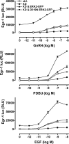
Effects of docking-domain mutants on GnRH-stimulated Egr-1 luciferase activity. Cells were transfected in 96-well plates and transduced with Ad mGnRHR and Ad Egr-1 luciferase. ERK knockdown (KD) was achieved by transfection with ERK1/2 siRNAs, and addback was with Ad WT ERK2-GFP or D319N ERK2-GFP as indicated (ctrl, control cells that received neither knockdown nor addback). After culture, these cells were stimulated for 4 h with the indicated concentrations of GnRHR, PDBu, or EGF. They were then processed for measurement of luciferase (luc) activity as described in Materials and Methods. The data shown are pooled from four separate experiments (mean ± sem, n = 2–4) each with duplicate or triplicate wells, and the figure shows Egr-1 luciferase activity in relative light units (RLU).
Fig. 7.
Fig. 7.
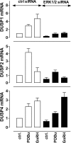
Effects of GnRH, PDBu, and ERK knockdown on nuclear-inducible DUSP mRNA. Cells were transfected in six-well plates with control siRNAs (ctrl) or ERK1/2 siRNAs and transduced with Ad mGnRHR before incubation for 120 min in control medium or with 1 μm PDBu or 1 μm GnRH, as indicated. Total RNA isolates were analyzed for relative levels of DUSP1, -2, or -4 mRNA by qPCR as described in Materials and Methods. The data shown are normalized values obtained from three separate experiments, each with duplicate readings (mean ± sem, n = 3). *, P < 0.05; **, P < 0.01, comparing control siRNA-transfected cells to ERK1/2 siRNA-transfected cells, using Student’s t test.
Fig. 8.
Fig. 8.
Effects of DUSP knockdown on DUSP induction by GnRH. Cells were transfected in six-well plates with control siRNAs (ctrl), DUSP2 siRNAs, or DUSP1 siRNA, transduced with Ad mGnRHR, and incubated for 120 min in control medium (−) or with 1 μm GnRH (+), as indicated. Total RNA isolates were analyzed for relative levels of DUSP1 or -4 mRNA by qPCR as described in Materials and Methods. The data shown are normalized values obtained from three separate experiments, each with duplicate readings (mean ± sem, n = 3). *, P < 0.05; **, P < 0.01, comparing control and GnRH-treated cells, using Student’s t test. In parallel experiments, effects of GnRH on DUSP2 mRNA levels were also assessed, and these were not measurably altered by knockdown of DUSP1 or -2 (not shown).
Fig. 9.
Fig. 9.
Effects of DUSP knockdown on ERK activation by GnRH, EGF, and PDBu. A and B, Cells were transfected in 96-well plates with ERK1/2 siRNAs and with either control siRNAs or with siRNAs targeting DUSPs 1, 2, and 4 (triple knockdown). They were also transduced with Ad ERK2-GFP and Ad mGnRHR before stimulation for the indicated periods with 1 μm GnRH. C and D, For the purpose of this comparison, the 60-min GnRH data from A and B have been plotted alongside previously published data from cells stimulated for 30 min with 10 nm EGF and or 1 μm PDBu (30 ). The data shown are pooled from six separate internal experiments (mean ± sem, n = 3–6), each with duplicate or triplicate wells. E, Cells were transfected in 96-well plates with ERK1/2 siRNAs and with either control siRNAs or with siRNAs targeting DUSPs 1, 2, and 4 (triple knockdown). They were also transduced with Ad ERK2-GFP, Ad mGnRHR, and Ad Egr-1 luciferase. After culture, they were stimulated for 4 h with GnRH (1 μm), PDBu (1 μm), or EGF (10 nm) and then processed for measurement of luciferase activity as described in Materials and Methods. The data shown are pooled from four separate experiments (mean ± sem, n = 2–4), each with duplicate or triplicate wells, and the figure shows Egr-1 luciferase activity in relative fluorescence units (RLU). **, P < 0.01; *, P < 0.05 compared with corresponding control values.
Fig. 10.
Fig. 10.
A screen for DUSP effects on GnRH signaling to ERK. Cells were transfected in 96-well plates with ERK1/2 siRNAs and with either control siRNAs or with siRNAs targeting each of the DUSPs indicated. They were also transduced with Ad ERK2-GFP and Ad mGnRHR before stimulation (0, 5, or 120 min) with 1 μm GnRH and then processed for imaging to determine whole-cell ppERK2 levels as well as the N:C ERK2-GFP ratio. Each of the indicated DUSPs are detectable by qPCR in these cells, and the siRNAs reduce expression of the appropriate transcripts by more than 70% (30 34 ). mRNA for DUSPs 13, 15, and 21 were not detectable in these cells, and we were unable to reduce DUSP8 expression by more than 30%, so these were excluded from the screen. None of the siRNAs that affected GnRH responses had any measurable effect on cell number or whole-cell ERK2-GFP levels, demonstrating that they do not have general toxic effects on cell survival, proliferation, transcription, or translation (supplemental table, published as supplemental data on The Endocrine Society’s Journals Online web site at http://mend.endojournals.org). One-way ANOVA revealed the siRNAs as significant variables (for each time point and endpoint), and statistically significant effects of the individual siRNA Smartpools are indicated (*, P < 0.05 by post hoc Bonferroni tests).

References

    1. Conn PM, Crowley Jr WF1994. Gonadotropin-releasing hormone and its analogs. Annu Rev Med 45:391–405 - PubMed
    1. Millar RP, Lu ZL, Pawson AJ, Flanagan CA, Morgan K, Maudsley SR2004. Gonadotropin-releasing hormone receptors. Endocr Rev 25:235–275 - PubMed
    1. Stojilkovic SS, Catt KJ1995. Expression and signal transduction pathways of gonadotropin-releasing hormone receptors. Recent Prog Horm Res 50:161–205 - PubMed
    1. Caunt CJ, Finch AR, Sedgley KR, McArdle CA2006. GnRH receptor signalling to ERK: kinetics and compartmentalization. Trends Endocrinol Metab 17:308–313 - PubMed
    1. Chen RH, Sarnecki C, Blenis J1992. Nuclear localization and regulation of erk- and rsk-encoded protein kinases. Mol Cell Biol 12:915–927 - PMC - PubMed

Publication types

MeSH terms

Substances