. 2009 May 1;179(9):772-81.
doi: 10.1164/rccm.200805-666OC. Epub 2009 Jan 29.

Resolution of allergic inflammation and airway hyperreactivity is dependent upon disruption of the T1/ST2-IL-33 pathway

Affiliations

Resolution of allergic inflammation and airway hyperreactivity is dependent upon disruption of the T1/ST2-IL-33 pathway

Jennifer Kearley et al. Am J Respir Crit Care Med. .

Abstract

Rationale: Although there have been numerous studies on the development of allergen-induced inflammation, the mechanisms leading to resolution of inflammation remain poorly understood. This represents an important consideration because failure to resolve allergen driven inflammation potentially leads to irreversible airway remodeling, characteristic of chronic asthma.

Objectives: We investigated the resolution of allergic inflammation and identified the factors responsible.

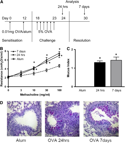
Methods: BALB/c and C57BL/6 mice were sensitized to ovalbumin and challenged through the airways to induce allergic inflammation. Mice were analyzed at 24 hours and 7 days after the final challenge.

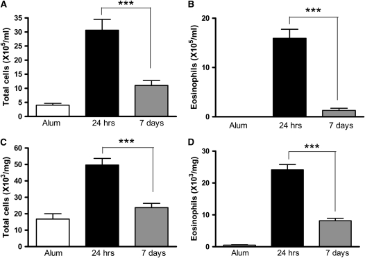
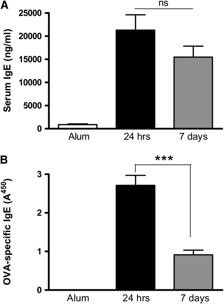
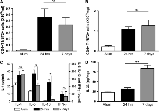
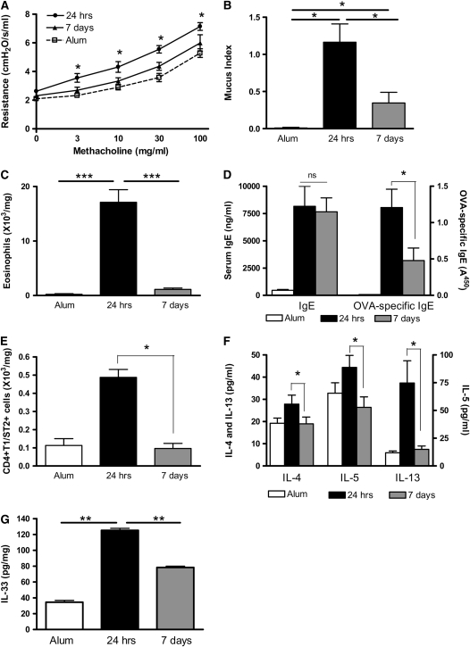
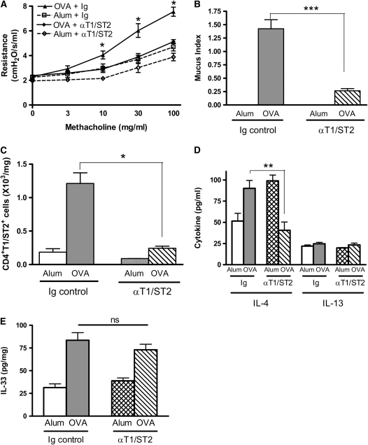
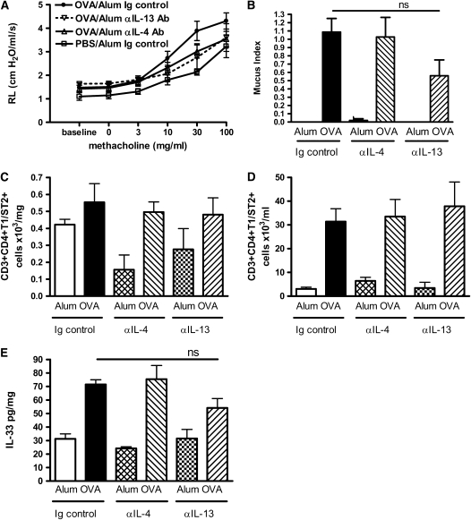
Measurements and main results: Airway hyperreactivity (AHR) and increased mucus production were present 7 days after the cessation of allergen challenge in BALB/c mice. Persisting AHR correlated with the continued presence of Th2 cells but not eosinophils in the lungs. The role of Th2 cells in maintaining AHR was confirmed using blocking antibodies against T1/ST2, IL-4, and IL-13 during the resolution period. Moreover, AHR in the "Th1 type" C57BL/6 mouse strain was resolved 1 week after allergen challenge, concomitant with clearance of Th2 cells from the lung. Expression of the T1/ST2 ligand, IL-33, also correlated with maintenance of AHR.

Conclusions: We have used blockade of Th2 function and strain differences to show for the first time that resolution of allergic inflammation and AHR may be dependent on the T1/ST2-IL-33 pathway and the presence of Th2 cells, suggesting they are necessary not only for the development of an allergic response but also for its maintenance.


Figures

<b>Figure 1.</b>
Figure 1.
(A) Airway hyperreactivity (AHR) and mucus production persist 1 week after allergen challenge in BALB/c mice. Airway inflammation was induced in BALB/c mice sensitized with ovalbumin (OVA) in alum on Days 0 and 12. One group of mice received alum instead of OVA/alum and served as the controls. All mice were subjected to daily aerosolized OVA challenge from Days 18 to 23 and were killed on Day 24, 24 hours after the final OVA challenge. A separate group of mice were killed on Day 30, 7 days after the final OVA challenge. (B) AHR was measured as lung resistance in BALB/c mice 24 hours and 7 days after the final OVA challenge in response to increasing concentrations of methacholine (3–100 mg/ml). Lungs were sectioned and stained with Periodic Acid-Schiff (PAS). (C) Sections were scored according to the criteria described in Methods. (D) Representative PAS-stained sections are shown for each group. Data are expressed as mean ± SEM (n = 6–16 mice/group from 1 to 3 independent experiments). *P < 0.05 compared with alum control mice.
<b>Figure 2.</b>
Figure 2.
Bronchoalveolar lavage (BAL) and lung leukocyte numbers decline after cessation of allergen challenge. (A and B) BAL and (C and D) lung tissue digest cells were isolated from BALB/c mice. Eosinophil numbers were determined by differential counts of BAL and lung digest cytospins. Data are expressed as mean ± SEM (n = 8 to 10 mice/group from two independent experiments). ***P < 0.001 comparing OVA-sensitized mice at 24 hours and 7 days after challenge.
<b>Figure 3.</b>
Figure 3.
Peripheral and local humoral responses decline after cessation of allergen challenge. Levels of (A) total IgE and (B) OVA-specific IgE were measured in the serum of BALB/c mice by ELISA. Data are expressed as mean ± SEM (n = 8–10 mice/group from two independent experiments). ***P < 0.001 comparing OVA-sensitized mice at 24 hours and 7 days after challenge.
<b>Figure 4.</b>
Figure 4.
T1/ST2+ Th2 cells and IL-4 persist in the airway lumen and lung of OVA-sensitized mice 1 week after allergen challenge. (A) BAL and (B) lung tissue digest cells were isolated from BALB/c mice. CD4+T1/ST2+ Th2 cell numbers were determined 24 hours or 7 days after the final OVA challenge by antibody staining and flow cytometric analysis. (C) IL-4, IL-5, and IL-13 levels were measured in BAL fluid by ELISA. (D) IL-33 levels were measured in lung tissue by ELISA. Values are expressed as mean ± SEM (n = 4–12 mice/group from two independent experiments).*P < 0.05 and **P < 0.01 comparing OVA-sensitized mice at 24 hours and 7 days after challenge.
<b>Figure 5.</b>
Figure 5.
Expression of Th2-attracting chemokines declines 1 week after the final OVA challenge. Levels of (A) CCL11/eotaxin, (B) CCL17/TARC, (C) CCL22/MDC, and (D) CCL1/TCA-3 were determined in BAL supernatant by ELISA in BALB/c mice 24 hours and 7 days after the final allergen challenge. Data are expressed as mean ± SEM (n = 8–10 mice/group from two independent experiments). *P < 0.05; **P < 0.01; ***P < 0.001.
<b>Figure 6.</b>
Figure 6.
AHR and Th2 cells do not persist in C57BL/6 mice 1 week after challenge. C57BL/6 mice were sensitized and challenged with OVA. (A) AHR was assessed by measuring lung resistance in response to increasing concentrations of methacholine (3–100 mg/ml). (B) Mucus secretion in PAS-stained lung sections were scored according to the criteria described in Methods. (C) Lung tissue eosinophils were determined by differential counts of lung digest cytospins. (D) Total serum IgE and OVA-specific IgE levels were measured by ELISA. (E) Lung tissue CD4+T1/ST2+ Th2 cells were quantified by antibody staining and flow cytometric analysis. (F) Th2 cytokine levels in BAL fluid and (G) IL-33 levels in lung tissue were measured by ELISA. Data are expressed as mean ± SEM (n = 8 to 10 mice/group from two independent experiments). *P < 0.05 and **P < 0.01 compared with alum control mice (A and B) or comparing OVA-sensitized mice at 24 hours and 7 days after challenge (CF).
<b>Figure 7.</b>
Figure 7.
Blockade of T1/ST2–IL-33 pathway reduces persistent AHR and mucus production in BALB/c mice. BALB/c mice were treated with anti-T1/ST2 antibody or rat Ig control antibody during the resolution phase (Days 25, 27, and 30). Airway inflammation and AHR were measured 7 days after the final OVA challenge. (A) AHR was assessed by measuring lung resistance in response to increasing concentrations of methacholine (3–100 mg/ml). (B) Mucus secretion was assessed in PAS-stained lung sections. (C) CD4+T1/ST2+ Th2 cell numbers were determined by antibody staining and flow cytometric analysis. (D) Th2 cytokine expression in BAL supernatant and (E) IL-33 in lung tissue were determined by ELISA. Data are expressed as mean ± SEM (n = 4–6 mice/group). *P < 0.05, **P < 0.01, and ***P < 0.001 compared with alum control mice (A) or comparing OVA-sensitized mice treated with anti-T1/ST2 antibody with those treated with control Ig (BE).
<b>Figure 8.</b>
Figure 8.
Blockade of IL-4 or IL-13 leads to partial resolution of AHR and mucus production. BALB/c mice were treated with anti–IL-4 antibody, anti–IL-13 antibody, or Ig control antibody during the resolution phase (Days 25, 27, and 30). Airway inflammation and AHR were measured 7 days after the final OVA challenge. (A) AHR was assessed by measuring lung resistance in response to increasing concentrations of methacholine (3–100 mg/ml). (B) Mucus secretion was assessed in PAS-stained lung sections. CD4+T1/ST2+ Th2 cell numbers in (C) lung tissue and (D) BAL were determined by antibody staining and flow cytometric analysis. (E) IL-33 in lung tissue were determined by ELISA. Data are expressed as mean ± SEM (n = 4–7 mice/group). *P < 0.05 and **P < 0.01 comparing anti–IL-13 antibody–treated OVA-challenged mice with Ig control–treated OVA-challenged mice. No statistically significant differences were measured between the OVA-challenged Ig control group compared with anti–IL-4 antibody–treated groups.

References

    1. Lloyd CM, Gonzalo JA, Coyle AJ, Gutierrez-Ramos JC. Mouse models of allergic airway disease. Adv Immunol 2001;77:263–295. - PubMed
    1. Bousquet J, Jeffery PK, Busse WW, Johnson M, Vignola AM. Asthma: from bronchoconstriction to airways inflammation and remodeling. Am J Respir Crit Care Med 2000;161:1720–1745. - PubMed
    1. Kay AB. The role of eosinophils in the pathogenesis of asthma. Trends Mol Med 2005;11:148–152. - PubMed
    1. Humbles AA, Lloyd CM, McMillan SJ, Friend DS, Xanthou G, McKenna E, Ghiran S, Gerard NP, Yu C, Orkin SH, et al. A critical role for eosinophils in allergic airways remodeling. Science 2004;305:1776–1779. - PubMed
    1. Lee JJ, Dimina D, Macias MP, Ochkur SI, McGarry MP, O'Neill KR, Protheroe C, Pero R, Nguyen T, Cormier SA, et al. Defining a link with asthma in mice congenitally deficient in eosinophils. Science 2004;305:1773–1776. - PubMed

Publication types

MeSH terms