

Aberrant heparan sulfate proteoglycan localization, despite normal exostosin, in central chondrosarcoma
- PMID: 19179614
- PMCID: PMC2665757
- DOI: 10.2353/ajpath.2009.080623
Aberrant heparan sulfate proteoglycan localization, despite normal exostosin, in central chondrosarcoma
Abstract
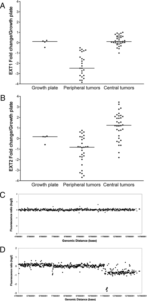
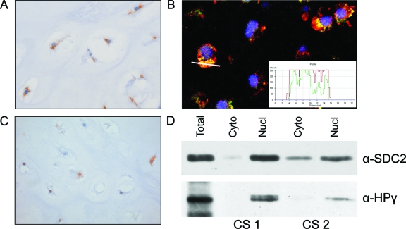
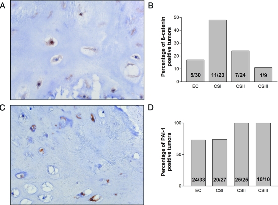
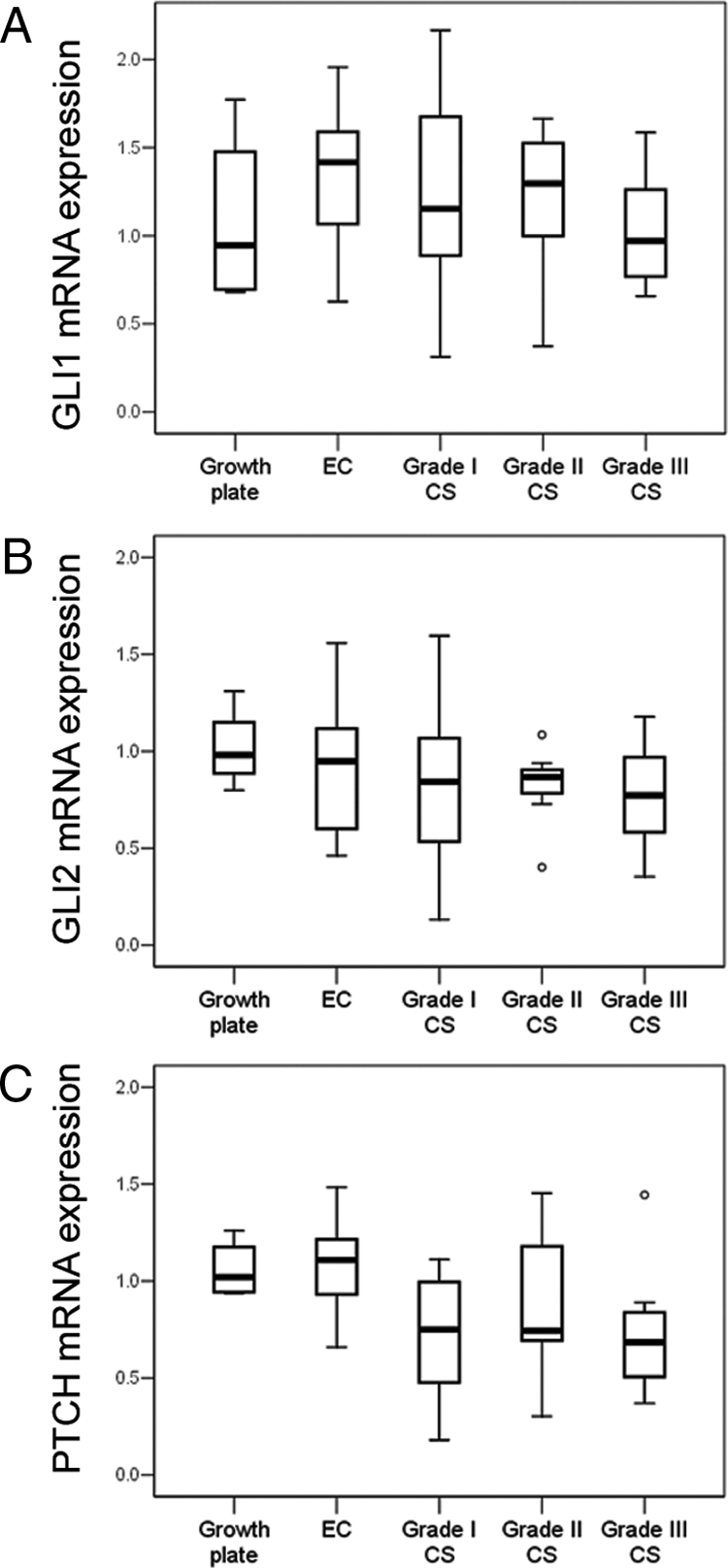
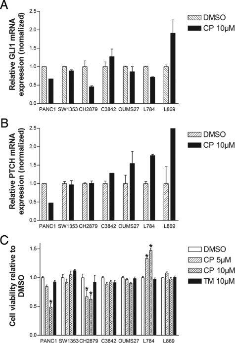
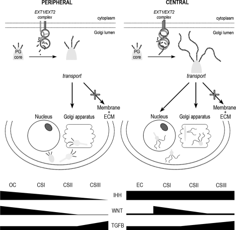
The tumor suppressor genes EXT1 and EXT2 are involved in the formation of multiple osteochondromas, which can progress to become secondary peripheral chondrosarcomas. The most common chondrosarcoma subtype is primary central chondrosarcoma, which occurs in the medullar cavity of bone. The EXT1/EXT2 protein complex is involved in heparan sulfate proteoglycan (HSPG) biosynthesis, which is important for signal transduction of Indian hedgehog (IHH), WNT, and transforming growth factor (TGF)-beta. The role of EXT and its downstream targets in central chondrosarcomas is currently unknown. EXT1 and EXT2 were therefore evaluated in central chondrosarcomas at both the DNA and mRNA levels. Immunohistochemistry was used to assess HSPG (CD44v3 and SDC2), WNT (beta-catenin), and TGF-beta (PAI-1 and phosphorylated Smad2) signaling, whereas IHH signaling was studied both by quantitative polymerase chain reaction and in vitro. mRNA levels of both EXT1 and EXT2 were normal in central chondrosarcomas; genomic alterations were absent in these regions and in 30 other HSPG-related genes. Although HSPGs were aberrantly located (CD44v3 in the Golgi and SDC2 in cytoplasm and nucleus), this was not caused by mutation. WNT signaling negatively correlated with increasing histological grade, whereas TGF-beta positively correlated with increasing histological grade. IHH signaling was active, and inhibition decreased cell viability in one of six cell lines. Our data suggest that, despite normal EXT in central chondrosarcomas, HSPGs and HSPG-dependent signaling are affected in both central and peripheral chondrosarcomas.
Figures
Similar articles
-
Decreased EXT expression and intracellular accumulation of heparan sulphate proteoglycan in osteochondromas and peripheral chondrosarcomas.J Pathol. 2007 Mar;211(4):399-409. doi: 10.1002/path.2127. J Pathol. 2007. PMID: 17226760
-
Secondary peripheral chondrosarcoma evolving from osteochondroma as a result of outgrowth of cells with functional EXT.Oncogene. 2012 Mar 1;31(9):1095-104. doi: 10.1038/onc.2011.311. Epub 2011 Aug 1. Oncogene. 2012. PMID: 21804604
-
Up-regulation of PTHrP and Bcl-2 expression characterizes the progression of osteochondroma towards peripheral chondrosarcoma and is a late event in central chondrosarcoma.Lab Invest. 2000 Dec;80(12):1925-34. doi: 10.1038/labinvest.3780202. Lab Invest. 2000. PMID: 11140704
-
Multiple osteochondromas.Orphanet J Rare Dis. 2008 Feb 13;3:3. doi: 10.1186/1750-1172-3-3. Orphanet J Rare Dis. 2008. PMID: 18271966 Free PMC article. Review.
-
Epiphyseal growth plate and secondary peripheral chondrosarcoma: the neighbours matter.J Pathol. 2012 Jan;226(2):219-28. doi: 10.1002/path.3003. Epub 2011 Nov 23. J Pathol. 2012. PMID: 21956842 Review.
Cited by
-
An Atlas of Heparan Sulfate Proteoglycans in the Postnatal Rat Lens.Invest Ophthalmol Vis Sci. 2021 Nov 1;62(14):5. doi: 10.1167/iovs.62.14.5. Invest Ophthalmol Vis Sci. 2021. PMID: 34730792 Free PMC article.
-
Spinal chondrosarcoma: a review.Sarcoma. 2011;2011:378957. doi: 10.1155/2011/378957. Epub 2011 Mar 8. Sarcoma. 2011. PMID: 21437176 Free PMC article.
-
No haploinsufficiency but loss of heterozygosity for EXT in multiple osteochondromas.Am J Pathol. 2010 Oct;177(4):1946-57. doi: 10.2353/ajpath.2010.100296. Epub 2010 Sep 2. Am J Pathol. 2010. PMID: 20813973 Free PMC article.
-
FOXM1 in sarcoma: role in cell cycle, pluripotency genes and stem cell pathways.Oncotarget. 2016 Jul 5;7(27):42792-42804. doi: 10.18632/oncotarget.8669. Oncotarget. 2016. PMID: 27074562 Free PMC article. Review.
-
IMP3, CDK4, MDM2 and β-catenin expression in Enchondroma and Central Chondrosarcoma: Diagnostic and prognostic utility.Clinics (Sao Paulo). 2024 Oct 4;79:100483. doi: 10.1016/j.clinsp.2024.100483. eCollection 2024. Clinics (Sao Paulo). 2024. PMID: 39368400 Free PMC article.
References
-
- Ahn J, Ludecke H-J, Lindow S, Horton WA, Lee B, Wagner MJ, Horsthemke B, Wells DE. Cloning of the putative tumour suppressor gene for hereditary multiple exostoses (EXT1). Nat Genet. 1995;11:137–143. - PubMed
-
- Wuyts W, Van Hul W, Wauters J, Nemtsova M, Reyniers E, Van Hul E, De Boulle K, De Vries BBA, Hendrickx J, Herrygers I, Bossuyt P, Balemans W, Fransen E, Vits L, Coucke P, Nowak NJ, Shows TB, Mallet L, Van den Ouweland AMW, McGaughran J, Halley DJJ, Willems P. Positional cloning of a gene involved in hereditary multiple exostoses. Hum Mol Genet. 1996;5:1547–1557. - PubMed
-
- Stickens D, Clines G, Burbee D, Ramos P, Thomas S, Hogue D, Hecht JT, Lovett M, Evans GA. The EXT2 multiple exostoses gene defines a family of putative tumour suppressor genes. Nat Genet. 1996;14:25–32. - PubMed
-
- Bertoni F, Bacchini P, Hogendoorn PCW. Chondrosarcoma. Fletcher CDM, Unni KK, Mertens F, editors. Lyon,: IARC Press,; World Health Organization Classification of Tumours. Pathology and Genetics of Tumours of Soft Tissue and Bone. 2002:pp 247–251.
-
- Mulder JD, Schütte HE, Kroon HM, Taconis WK. Amsterdam: Elsevier,; Radiologic Atlas of Bone Tumors. 1993:pp 139–171.
Publication types
MeSH terms
Substances
LinkOut - more resources
Full Text Sources
Medical
Miscellaneous
