

Shared circuitry: developmental signaling cascades regulate both embryonic and adult coronary vasculature
- PMID: 19179669
- PMCID: PMC2757940
- DOI: 10.1161/CIRCRESAHA.108.191239
Shared circuitry: developmental signaling cascades regulate both embryonic and adult coronary vasculature
Abstract
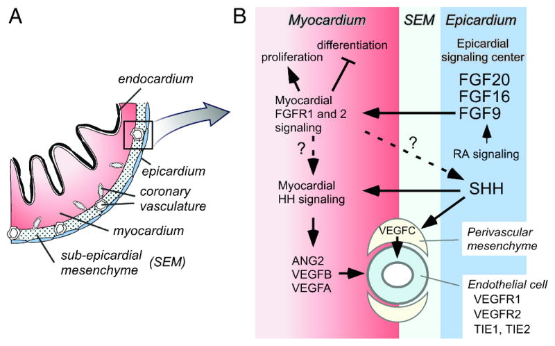
Ischemic heart disease is the most common cause of heart failure and is among the leading causes of mortality worldwide. Therapies used for the treatment of this disease aim to restore blood flow to severely narrowed or occluded coronary arteries by either catheter-based or surgical means. Although these strategies prove efficacious for many patients, a substantial number of individuals fail to improve following these procedures. Recently, a noninvasive strategy has been proposed, focusing on the use of endogenous growth factors that trigger the growth of new coronary arteries. Using the developing heart as a model, several groups have identified some of the key pathways that not only govern the development of the coronary vascular system but also promote the growth of the adult coronary vasculature. Here, we review the major morphological events and signaling cascades that mediate the formation of the coronary vasculature in the embryo. We further describe the mechanism by which many of these same pathways also regulate the adult coronary vasculature and their potential use in the treatment of ischemic heart disease.
Figures


Similar articles
-
Rebuilding the coronary vasculature: hedgehog as a new candidate for pharmacologic revascularization.Trends Cardiovasc Med. 2007 Apr;17(3):77-83. doi: 10.1016/j.tcm.2007.01.002. Trends Cardiovasc Med. 2007. PMID: 17418368 Free PMC article. Review.
-
Epicardial-myocardial signaling directing coronary vasculogenesis.Circ Res. 2010 Mar 19;106(5):818-32. doi: 10.1161/CIRCRESAHA.109.209197. Circ Res. 2010. PMID: 20299672 Free PMC article. Review.
-
Fibroblast growth factor signals regulate a wave of Hedgehog activation that is essential for coronary vascular development.Genes Dev. 2006 Jun 15;20(12):1651-66. doi: 10.1101/gad.1411406. Genes Dev. 2006. PMID: 16778080 Free PMC article.
-
Cellular origin and developmental program of coronary angiogenesis.Circ Res. 2015 Jan 30;116(3):515-30. doi: 10.1161/CIRCRESAHA.116.305097. Circ Res. 2015. PMID: 25634974 Free PMC article. Review.
-
Fibroblast growth factors and Hedgehogs: at the heart of the epicardial signaling center.Trends Genet. 2008 Jan;24(1):33-40. doi: 10.1016/j.tig.2007.10.007. Epub 2007 Dec 3. Trends Genet. 2008. PMID: 18054407 Review.
Cited by
-
Myocyte proliferation in the developing heart.Dev Dyn. 2011 Jun;240(6):1322-34. doi: 10.1002/dvdy.22650. Epub 2011 May 2. Dev Dyn. 2011. PMID: 21538685 Free PMC article. Review.
-
Microvascular repair: post-angiogenesis vascular dynamics.Microcirculation. 2012 Nov;19(8):676-95. doi: 10.1111/j.1549-8719.2012.00207.x. Microcirculation. 2012. PMID: 22734666 Free PMC article. Review.
-
Hedgehog-mediated regulation of PPARγ controls metabolic patterns in neural precursors and shh-driven medulloblastoma.Acta Neuropathol. 2012 Apr;123(4):587-600. doi: 10.1007/s00401-012-0968-6. Epub 2012 Mar 11. Acta Neuropathol. 2012. PMID: 22407012 Free PMC article.
-
Coronary arteries form by developmental reprogramming of venous cells.Nature. 2010 Mar 25;464(7288):549-53. doi: 10.1038/nature08873. Nature. 2010. PMID: 20336138 Free PMC article.
-
p38 MAPK priming boosts VSMC proliferation and arteriogenesis by promoting PGC1α-dependent mitochondrial dynamics.Sci Rep. 2022 Apr 8;12(1):5938. doi: 10.1038/s41598-022-09757-x. Sci Rep. 2022. PMID: 35396524 Free PMC article.
References
-
- Caines AE, Massad MG, Kpodonu J, Rebeiz AG, Evans A, Geha AS. Outcomes of coronary artery bypass grafting versus percutaneous coronary intervention and medical therapy for multivessel disease with and without left ventricular dysfunction. Cardiology. 2004;101:21–28. - PubMed
-
- Syed IS, Sanborn TA, Rosengart TK. Therapeutic angiogenesis: a biologic bypass. Cardiology. 2004;101:131–143. - PubMed
-
- Saraste A, Nekolla S, Schwaiger M. Contrast-enhanced magnetic resonance imaging in the assessment of myocardial infarction and viability. J Nucl Cardiol. 2008;15:105–117. - PubMed
-
- House SL, Bolte C, Zhou M, Doetschman T, Klevitsky R, Newman G, Schultz Jel J. Cardiac-specific overexpression of fibroblast growth factor-2 protects against myocardial dysfunction and infarction in a murine model of low-flow ischemia. Circulation. 2003;108:3140–3148. - PubMed
-
- Landau C, Jacobs AK, Haudenschild CC. Intrapericardial basic fibroblast growth factor induces myocardial angiogenesis in a rabbit model of chronic ischemia. Am Heart J. 1995;129:924–931. - PubMed
Publication types
MeSH terms
Substances
Grants and funding
LinkOut - more resources
Full Text Sources
