. 2009;4(2):206-23.
doi: 10.1038/nprot.2008.227.

Recombineering: a homologous recombination-based method of genetic engineering

Affiliations

Recombineering: a homologous recombination-based method of genetic engineering

Shyam K Sharan et al. Nat Protoc. 2009.

Abstract

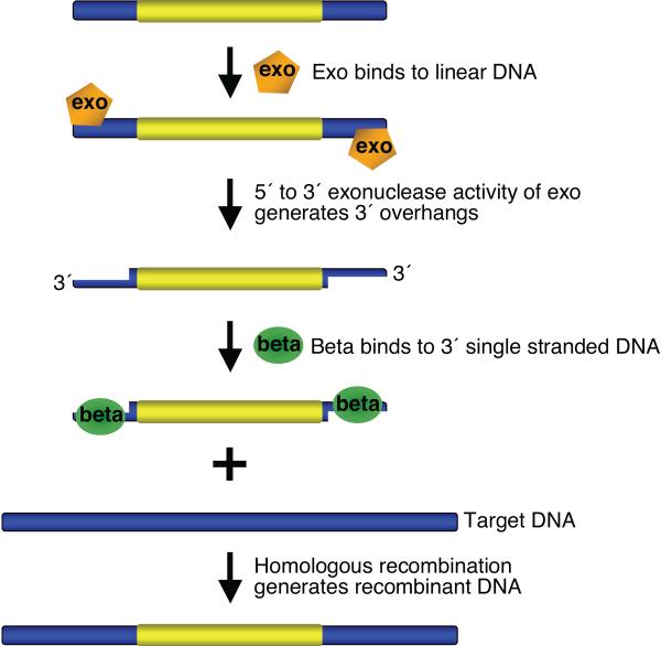
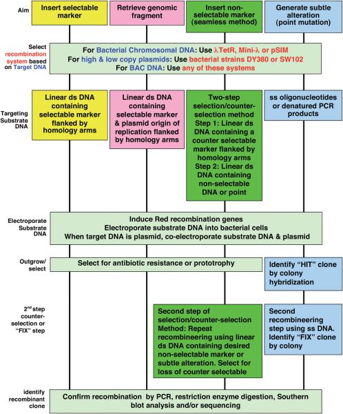
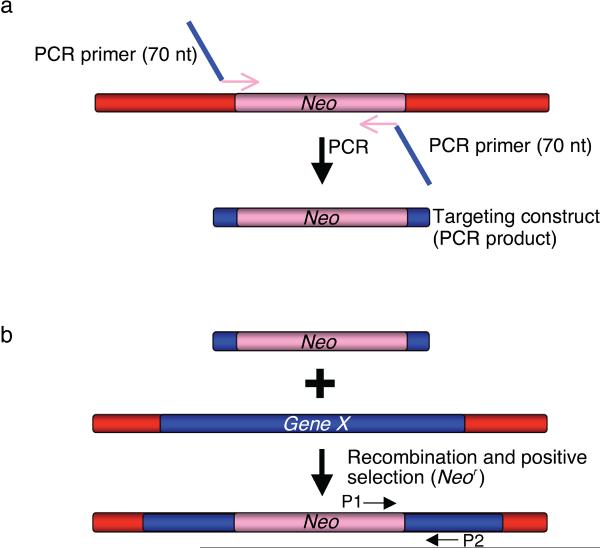
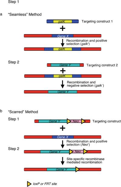
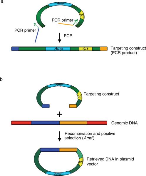
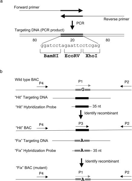
Recombineering is an efficient method of in vivo genetic engineering applicable to chromosomal as well as episomal replicons in Escherichia coli. This method circumvents the need for most standard in vitro cloning techniques. Recombineering allows construction of DNA molecules with precise junctions without constraints being imposed by restriction enzyme site location. Bacteriophage homologous recombination proteins catalyze these recombineering reactions using double- and single-stranded linear DNA substrates, so-called targeting constructs, introduced by electroporation. Gene knockouts, deletions and point mutations are readily made, gene tags can be inserted and regions of bacterial artificial chromosomes or the E. coli genome can be subcloned by gene retrieval using recombineering. Most of these constructs can be made within about 1 week's time.


Figures

Figure 1
Figure 1. Overview of bacteriophage l recombination system used for recombineering
Exo has a 5′ to 3′ dsDNA exonuclease activity, which can generate 3′ overhangs on linear DNA. Beta binds the single stranded DNA (3′ overhangs), promotes ss-annealing and generates recombinant DNA. An additional protein, Gam (not shown here), which prevents RecBCD nuclease from degrading double-strand linear DNA fragments, is also required for dsDNA recombineering.
Figure 2
Figure 2. A flow-chart of recombineering procedures
Schematic representation of various steps involved in recombineering. An appropriate system should be selected based on the choice of target DNA. The type of substrate DNA depends upon the choice of method used. The outgrowth procedures and methods to identify the recombinant clone are based on the use a selectable marker, selection/counter-selection method or lack of any selectable marker in the substrate DNA.
Figure 3
Figure 3. Insertion of a selectable marker by recombineering
(a) Targeting construct can be generated by PCR to introduce the region of homology (in blue) and a selectable marker (e.g. Neo, Kanamycin/Neomycin resistance gene). The PCR primers used to generate the targeting construct are 70-mer oligonucleotides with 50 nucleotides corresponding to the target site (e.g. Gene X, in blue) sequence to introduce the homology arm and 20 bases from the ends of the selectable marker (Neo, in pink). (b) The targeting construct is electroporated into the bacterial cells that are induced to express the phage recombination genes. Recombinant clones are selected as kanamycin resistant colonies and confirmed with PCR using test primers P1 and P2.
Figure 4
Figure 4. Insertion of a non-selectable DNA fragment by recombineering
(a) “Seamless” method to insert non-selectable DNA fragment makes use of selectable markers that can be used for positive as well as negative selection (e.g. galK). In this two-step method, first the selectable galK marker is targeted to the site (Gene X) where the non-selectable DNA fragment (Gene Y) is to be inserted. In the second step, a targeting construct containing the non-selectable DNA fragment flanked by the same 50 bp of homology to the target site is electroporated into Gal+ bacterial cells containing the recombinant DNA from step 1. Clones in which the Gene Y DNA fragment is correctly targeted are counter-selected for loss of the galK gene. (b) The scarred method: this method targets both the selected (Neo) and non-selected (Gene Y) DNAs jointly. In step 1, the non-selectable DNA fragment (Gene Y) is introduced along with a selectable marker, Neo, which is flanked by loxP or FRT sites. Recombinants are selected for the presence of Neo. In step 2, Neo is deleted by site-specific recombinase mediated recombination (Cre for loxP sites and Flp for FRT sites). Unlike the “seamless” method, a single loxP or FRT site is retained after recombination.
Figure 5
Figure 5. Subcloning DNA fragments from genomic DNA
(a) To subclone or retrieve a genomic DNA fragment, generate a targeting construct by PCR. Each primer consists of 50 bases from the end of the genomic DNA that needs to be subcloned (blue and orange) and 20 bases from the plasmid sequence (in green) flanking the region between the origin of replication (ori) and an antibiotic resistance marker (Amp, ampicillin resistance gene). A linear plasmid DNA is used as template for PCR. (b) The linear targeting construct (PCR product) is electroporated into the bacterial cells that are induced to express the phage recombination genes. Recombination between the targeting construct and the genomic DNA results in the formation of a circular plasmid by gap repair. The circular plasmid contains the desired DNA fragment.
Figure 6
Figure 6. Two-step “hit & fix” method to generate subtle mutations using single stranded short PCR product or oligonucleotides as targeting vector
(a) The single-stranded oligonucleotides containing 160 bases of homology and 20 unique bases are generated by using two 100-mer oligonucleotides in a PCR reaction. The two 100-mer oligonucleotides have 20 complementary bases (in this case the 20 bp contains restriction sites BamHI, EcoRV, XhoI) at the 3′ end. The 180 bp PCR product can be denatured to obtain single-stranded oligonucleotides that can be used as targeting construct. (b) Schematic representation of the two steps involved in “hit and fix” method to generate subtle alterations (e.g. G to A) without the use of a selectable marker. In step 1, a 180-mer single-strand oligonucleotide is used to replace 20 nucleotides (gray box) around the target site with 20 heterologous nucleotides (black box). Recombinants can be identified by colony hybridization using an end-labeled 35-mer oligonuclotide that can specifically anneal only to the recombinant DNA. A primer set specific for the heterologous “hit” sequence (P3 and P2) can be used to confirm the presence of recombinant clones by PCR. A second primer set (P1 and P2) can be used as a control to amplify only the non-recombinant DNA. Generation of a correct recombinant clone can be confirmed by digesting with BamHI, EcoRV or XhoI the PCR product (~300–500 bp) of primers P2 and P4. In step 2, the 20 nucleotides are restored to the original sequence, except for the desired mutation. Such clones can be identified by colony hybridization using a 35-mer oligonucleotide as probe and further confirmed by PCR amplification using primers P1 and P2, by testing for loss of the restriction sites inserted in step 1, by digesting the PCR product of primers P2 and P4, and by sequencing.
Figure 7
Figure 7. Schematic representation of λ phage constructs used for recombineering
(a) The λ prophage with some of its genes is shown integrated in the bacterial chromosome. It is adjacent to the biotin genes, bioAB. Bacterial DNA is shown as a blue bar; phage DNA is a red bar; and a region containing a deletion and/or a substitution is a white bar. The complete λ prophage is flanked by its attachments site att, where integration occurred. The int and xis genes located in the pL operon encode functions to integrate and excise the phage DNA into and out of the bacterial chromosome. Two operons with their promoters pL and pR are indicated. Transcription of the promoters is controlled by the λ CI857 repressor. This repressor is temperature sensitive in that it is active and represses the promoter at 30–32°C but is inactive at 42°C allowing transcription. The N functions as an anti-transcription terminator and prevents RNA polymerase termination of pL transcripts at terminators tL1, tL2, and tL3. The red genes exo, bet, and gam encoding the homologous recombination functions are also in the pL operon. The kil gene is adjacent to gam and when expressed for over 1 hr kills the bacterial cell . The replication genes O and P are in the pR operon. Cro functions as a partial repressor of the pL and pR operons when CI is inactive at 42°C. The lysis and structural genes, SRA-J, are shown located beyond their regulator Q. The λ TetR phage used for recombineering is identical to the above complete prophage with the exception of five changes. The replication gene P has an amber mutation, the cro gene also has an amber mutation, there is an added mutation ind1 in the cI857 repressor gene, and the tetA gene encoding tetracycline resistance replaces rex. (b) The defective prophages in DY380, SW102, and the HME strains have the lytic pR operon deleted from cro through the bioA genes as shown in brackets. The right att site is also deleted preventing any excision of this prophage. In DY380 and SW102 cro through bioA is replaced by the tetracycline resistance cassette, tetRA. (c) The mini-λ phage DNA is shown with the lytic genes cro through J deleted. In different constructs of the mini λ, the cro-J region is replaced with various drug resistant cassettes. Mini-λ has both att sites plus int and xis, which allows for its integration and excision. (d) The λ segment on the pSIM plasmids is shown. The plasmid backbone is not shown. The genes from cro to att are removed as well as genes beyond tL3 including int and xis. The red genes are connected directly to pL by a deletion that removes kil through N. The drug resistant marker characteristic of each SIM plasmid replaces the rex gene adjacent to cI857. The basic features are conserved in all of these Red expression constructs. Red is left under the native phage controlling elements for optimal expression and regulation. The pL promoter drives gene expression and is controlled by the temperature sensitive but renaturable λ CI857 repressor. In all, the cro gene is inactive to maximize pL expression, and the replication genes are absent or inactive to prevent lethal effects on the cell.

References

    1. Copeland NG, Jenkins NA, Court DL. Recombineering: a powerful new tool for mouse functional genomics. Nat. Rev. Genet. 2001;2:769–779. - PubMed
    1. Ellis HM, Yu D, DiTizio T, Court DL. High efficiency mutagenesis, repair, and engineering of chromosomal DNA using single-stranded oligonucleotides. Proc. Natl. Acad. Sci. U. S. A. 2001;98:6742–6746. - PMC - PubMed
    1. Yu D, et al. An efficient recombination system for chromosome engineering in Escherichia coli. Proc. Natl. Acad. Sci. U. S. A. 2000;97:5978–5983. - PMC - PubMed
    1. Zhang Y, Buchholz F, Muyrers JP, Stewart AF. A new logic for DNA engineering using recombination in Escherichia coli. Nat. Genet. 1998;20:123–128. - PubMed
    1. Zhang Y, Muyrers JP, Testa G, Stewart AF. DNA cloning by homologous recombination in Escherichia coli. Nat. Biotechnol. 2000;18:1314–1317. - PubMed

Publication types

MeSH terms