

Poisson-Nernst-Planck models of nonequilibrium ion electrodiffusion through a protegrin transmembrane pore
- PMID: 19180178
- PMCID: PMC2614469
- DOI: 10.1371/journal.pcbi.1000277
Poisson-Nernst-Planck models of nonequilibrium ion electrodiffusion through a protegrin transmembrane pore
Abstract
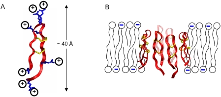
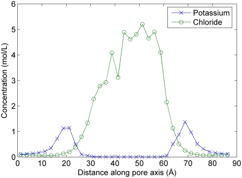
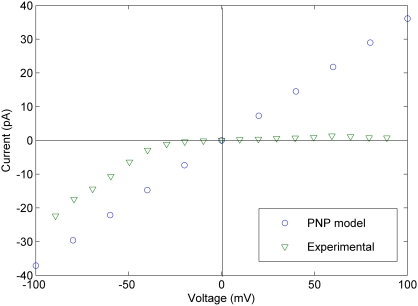
Protegrin peptides are potent antimicrobial agents believed to act against a variety of pathogens by forming nonselective transmembrane pores in the bacterial cell membrane. We have employed 3D Poisson-Nernst-Planck (PNP) calculations to determine the steady-state ion conduction characteristics of such pores at applied voltages in the range of -100 to +100 mV in 0.1 M KCl bath solutions. We have tested a variety of pore structures extracted from molecular dynamics (MD) simulations based on an experimentally proposed octomeric pore structure. The computed single-channel conductance values were in the range of 290-680 pS. Better agreement with the experimental range of 40-360 pS was obtained using structures from the last 40 ns of the MD simulation, where conductance values range from 280 to 430 pS. We observed no significant variation of the conductance with applied voltage in any of the structures that we tested, suggesting that the voltage dependence observed experimentally is a result of voltage-dependent channel formation rather than an inherent feature of the open pore structure. We have found the pore to be highly selective for anions, with anionic to cationic current ratios (I(Cl-)/I(K+)) on the order of 10(3). This is consistent with the highly cationic nature of the pore but surprisingly in disagreement with the experimental finding of only slight anionic selectivity. We have additionally tested the sensitivity of our PNP model to several parameters and found the ion diffusion coefficients to have a significant influence on conductance characteristics. The best agreement with experimental data was obtained using a diffusion coefficient for each ion set to 10% of the bulk literature value everywhere inside the channel, a scaling used by several other studies employing PNP calculations. Overall, this work presents a useful link between previous work focused on the structure of protegrin pores and experimental efforts aimed at investigating their conductance characteristics.
Conflict of interest statement
The authors have declared that no competing interests exist.
Figures
Similar articles
-
Ion permeation and selectivity of OmpF porin: a theoretical study based on molecular dynamics, Brownian dynamics, and continuum electrodiffusion theory.J Mol Biol. 2002 Sep 27;322(4):851-69. doi: 10.1016/s0022-2836(02)00778-7. J Mol Biol. 2002. PMID: 12270719
-
Ion permeation through the alpha-hemolysin channel: theoretical studies based on Brownian dynamics and Poisson-Nernst-Plank electrodiffusion theory.Biophys J. 2004 Oct;87(4):2299-309. doi: 10.1529/biophysj.104.044008. Biophys J. 2004. PMID: 15454431 Free PMC article.
-
Optimization of 3D Poisson-Nernst-Planck model for fast evaluation of diverse protein channels.Proteins. 2013 Oct;81(10):1802-22. doi: 10.1002/prot.24326. Epub 2013 Aug 19. Proteins. 2013. PMID: 23720356
-
Theoretical simulation of the ion current rectification (ICR) in nano-pores based on the Poisson-Nernst-Planck (PNP) model.Phys Chem Chem Phys. 2014 Jan 7;16(1):23-32. doi: 10.1039/c3cp51712h. Phys Chem Chem Phys. 2014. PMID: 24253284 Review.
-
Emerging issues of connexin channels: biophysics fills the gap.Q Rev Biophys. 2001 Aug;34(3):325-472. doi: 10.1017/s0033583501003705. Q Rev Biophys. 2001. PMID: 11838236 Review.
Cited by
-
Collective diffusion model for ion conduction through microscopic channels.Biophys J. 2013 Jan 22;104(2):368-76. doi: 10.1016/j.bpj.2012.11.3826. Biophys J. 2013. PMID: 23442858 Free PMC article.
-
Computational studies of protegrin antimicrobial peptides: a review.Peptides. 2011 Jan;32(1):188-201. doi: 10.1016/j.peptides.2010.10.006. Epub 2010 Oct 12. Peptides. 2011. PMID: 20946928 Free PMC article. Review.
-
A Stabilized Finite Element Method for Modified Poisson-Nernst-Planck Equations to Determine Ion Flow Through a Nanopore.Commun Comput Phys. 2014 Jan;15(1):10.4208/cicp.101112.100413a. doi: 10.4208/cicp.101112.100413a. Commun Comput Phys. 2014. PMID: 24363784 Free PMC article.
-
Modeling the electric potential across neuronal membranes: the effect of fixed charges on spinal ganglion neurons and neuroblastoma cells.PLoS One. 2014 May 6;9(5):e96194. doi: 10.1371/journal.pone.0096194. eCollection 2014. PLoS One. 2014. PMID: 24801682 Free PMC article.
-
Relative free energy of binding between antimicrobial peptides and SDS or DPC micelles.Mol Simul. 2009 Sep;35(10-11):986-997. doi: 10.1080/08927020902902742. Mol Simul. 2009. PMID: 21113423 Free PMC article.
References
-
- Zasloff M. Antimicrobial peptides of multicellular organisms. Nature. 2002;415:389–395. - PubMed
-
- Brogden KA. Antimicrobial peptides: pore formers or metabolic inhibitors in bacteria? Nat Rev Microbiol. 2005;3:238–250. - PubMed
-
- Hancock RE. Peptide antibiotics. Lancet. 1997;349:418–422. - PubMed
-
- Wu M, Maier E, Benz R, Hancock RE. Mechanism of interaction of different classes of cationic antimicrobial peptides with planar bilayers and with the cytoplasmic membrane of Escherichia coli. Biochemistry. 1999;38:7235–7242. - PubMed
-
- Yeaman MR, Yount NY. Mechanisms of antimicrobial peptide action and resistance. Pharmacol Rev. 2003;55:27–55. - PubMed
Publication types
MeSH terms
Substances
Grants and funding
LinkOut - more resources
Full Text Sources
Miscellaneous
