Case Reports
. 2009 Feb;60(2):584-91.
doi: 10.1002/art.24221.

Molecular framework for response to imatinib mesylate in systemic sclerosis

Affiliations
Case Reports

Molecular framework for response to imatinib mesylate in systemic sclerosis

Lorinda Chung et al. Arthritis Rheum. 2009 Feb.

Abstract

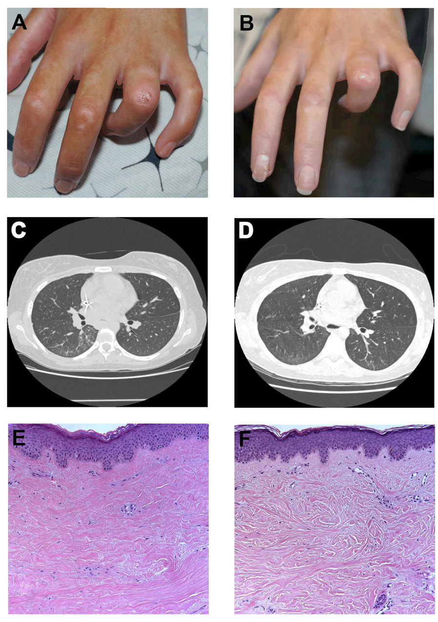
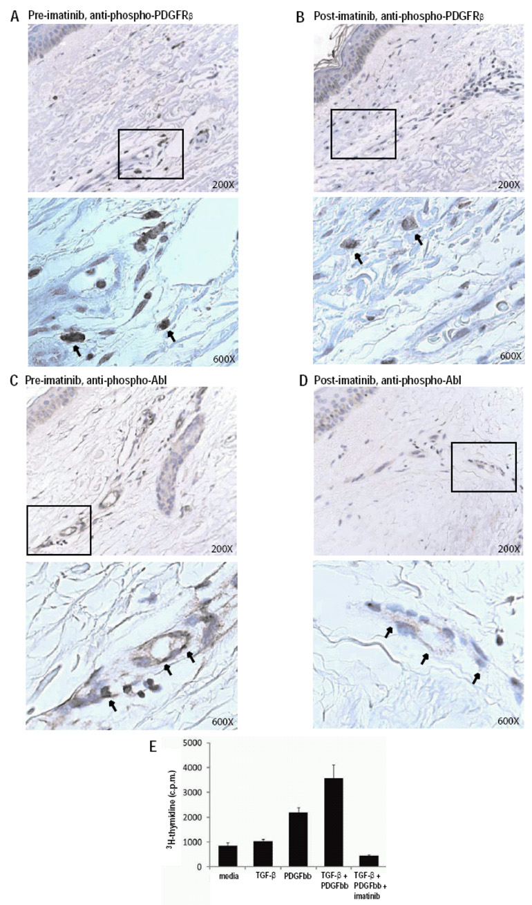
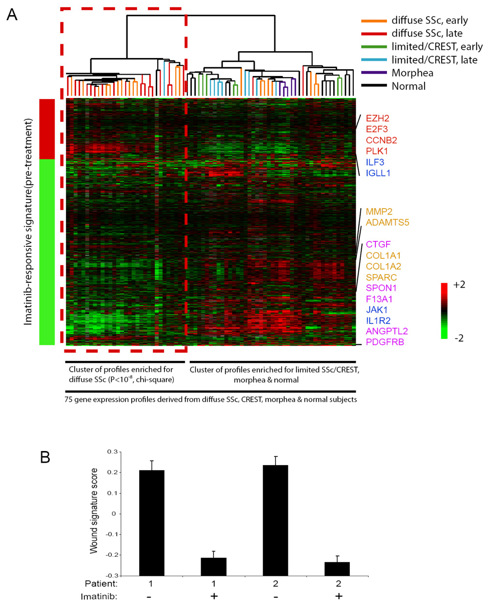
Systemic sclerosis (SSc) is an autoimmune disease in which the tyrosine kinases platelet-derived growth factor receptor (PDGFR) and Abl are hypothesized to contribute to the fibrosis and vasculopathy of the skin and internal organs. Herein we describe 2 patients with early diffuse cutaneous SSc (dcSSc) who experienced reductions in cutaneous sclerosis in response to therapy with the tyrosine kinase inhibitor imatinib mesylate. Immunohistochemical analyses of skin biopsy specimens demonstrated reductions of phosphorylated PDGFRbeta and Abl with imatinib therapy. By gene expression profiling, an imatinib-responsive signature specific to dcSSc was identified (P < 10(-8)). The response of these patients and the findings of the analyses suggest that PDGFRbeta and Abl play critical, synergistic roles in the pathogenesis of SSc, and that imatinib targets a gene expression program that is frequently dysregulated in dcSSc.


Figures

Figure 1
Figure 1. Effect of imatinib on digital ulcers, interstitial lung disease, and collagen architecture in a patient with SSc
(A) Digital ulcer located over the left fourth proximal interphalangeal joint prior to imatinib therapy. (B) Healing of digital ulcer after 3 months of imatinib therpy. (C) HRCT of the chest prior to imatinib therapy demonstrates patchy infiltrates associated with ground glass opacities in the bilateral lower lobes. (D) HRCT after 3 months of imatinib therapy shows resolution of ground glass opacities. (E) Hematoxylin and eosin stained skin biopsy from the right arm taken prior to imatinib therapy shows dense, eosinophilic, tightly packed collagen bundles of the papillary and reticular dermis with an average dermal thickness of 2.81 mm (Magnification 100×). (F) Skin biopsy after 3 months of imatinib taken within 1 cm of initial biopsy shows normalization of collagen architecture, with loose spacing and thinning of collagen bundles and an average dermal thickness of 2.31 mm.
Figure 2
Figure 2. Imatinib reduces PDGFRβ and Abl activation in SSc skin and function in SSc fibroblasts
(A–D) Immunohistochemical staining of serial skin biopsy samples obtained pretreatment (A,C) and one month following the initiation of imatinib treatment (B,D) with anti-phospho-PDGFRβ (A,B) and anti-phospho-Abl (C,D) antibodies. Boxed areas of upper panels (200× magnification), are presented at higher magnification in their corresponding lower panels (600×). Results are representative of those obtained from multiple sections from two independent patients. Phospho-PDGFRβ was observed in interstitial fibroblasts as well as perivascular spindle-like cells and some cells resembling mast cells. Phospho-Abl was observed in endothelial cells in small vessels and in scattered dermal fibroblasts. (E) Stimulation of a SSc fibroblast line with PDGF (10 ng/ml), TGF-β (0.5 ng/ml), PDGF + TGF-β, or PDGF + TGF-β+ imatinib (1 µM). Proliferation was quantitated after 48 hours by 3H-thymidine incorporation (Y axis). Results are representative of experiments performed on two independent SSc fibroblast lines, and similar results were obtained with normal fibroblast lines.
Figure 3
Figure 3. An imatinib-responsive signature is present in most diffuse SSc
(A) The imatinib-responsive signature was determined by applying Significance Analysis of Microarrays (SAM) to identify mRNA that exhibited statistically significant changes in their levels in pre-treatment as compared to post-treatment skin biopsy samples derived from the two imatinib-treated SSc patients. SAM identified 1050 genes that were changed by imatinib therapy in both patients (FDR<0.001), and this imatinib responsive signature is represented by the bar to the left of the heatmap image (red represents an increase, and green a decrease, in mRNA expression post-treatment; the genes comprising the imatinib responsive signature are presented in Supplemental Table 1). The imatinib-responsive genes were then used to organize via unsupervised hierarchical clustering the 75 gene expression profiles derived from skin biopsies from SSc, limited SSc/CREST, morphea and health control patients contained in a database. The results of the hierarchical clustering are presented as a heatmap, with each column representing the mRNA profile of a sample, and rows representing the genes present in the imatinib responsive signature. Unsupervised hierarchical clustering revealed two distinct clusters, with the imatinib-responsive gene expression pattern being similar to one of the clusters, and this cluster being highly enriched for diffuse SSc samples (29 out of the 31 gene expression profiles contained in this cluster are from diffuse SSc, P<10−8, chi-square). This cluster of gene expression profiles derived from most of the diffuse SSc samples exhibited a pattern of gene activation and repression concordant with the imatinib-responsive signature, including alterations in the expression of genes involved in cell proliferation (red), immune signaling (blue), matrix remodeling (tan), and growth factor signaling (pink) (indicated to the right of the heatmap). The other cluster contained most of the profiles derived from limited/CREST, morphea and normal subjects, and the gene expression profiles from these patients did not exhibit the imatinib-responsive signature (this cluster contains 44 gene expression profiles, including 14 from normal skin, 15 from limited SSc/CREST, 5 from morphea, and 10 from diffuse SSc). (B) Reduction in the wound signature by imatinib in two patients with SSc. Replicate array analysis was performed for each sample; mean ± standard deviation is shown.

References

    1. Baroni SS, Santillo M, Bevilacqua F, Luchetti M, Spadoni T, Mancini M, et al. Stimulatory autoantibodies to the PDGF receptor in systemic sclerosis. N Engl J Med. 2006;354(25):2667–2676. - PubMed
    1. Ludwicka A, Ohba T, Trojanowska M, Yamakage A, Strange C, Smith EA, et al. Elevated levels of platelet derived growth factor and transforming growth factor-beta 1 in bronchoalveolar lavage fluid from patients with scleroderma. J Rheumatol. 1995;22(10):1876–1883. - PubMed
    1. Yamakage A, Kikuchi K, Smith EA, LeRoy EC, Trojanowska M. Selective upregulation of platelet-derived growth factor alpha receptors by transforming growth factor beta in scleroderma fibroblasts. J Exp Med. 1992;175(5):1227–1234. - PMC - PubMed
    1. Schermuly RT, Dony E, Ghofrani HA, Pullamsetti S, Savai R, Roth M, et al. Reversal of experimental pulmonary hypertension by PDGF inhibition. J Clin Invest. 2005;115(10):2811–2821. - PMC - PubMed
    1. Soria A, Cario-Andre M, Lepreux S, Rezvani HR, Pasquet JM, Pain C, et al. The effect of imatinib (Glivec) on scleroderma and normal dermal fibroblasts: a preclinical study. Dermatology. 2008;216(2):109–117. - PubMed

Publication types

MeSH terms