. 2009 Feb;60(2):492-500.
doi: 10.1002/art.24225.

Increased expression of the Akt/PKB inhibitor TRB3 in osteoarthritic chondrocytes inhibits insulin-like growth factor 1-mediated cell survival and proteoglycan synthesis

Affiliations

Increased expression of the Akt/PKB inhibitor TRB3 in osteoarthritic chondrocytes inhibits insulin-like growth factor 1-mediated cell survival and proteoglycan synthesis

John D Cravero et al. Arthritis Rheum. 2009 Feb.

Abstract

Objective: The chondrocyte response to insulin-like growth factor 1 (IGF-1) is reduced with aging and in osteoarthritis (OA). IGF-1 signals through the phosphatidylinositol 3-kinase/Akt pathway. TRB3, a tribbles homolog, has been shown to inhibit IGF-1-mediated activation of Akt in HEK 293 cells. This study was undertaken to determine if TRB3 is expressed in chondrocytes, and whether the chondrocyte response to IGF-1 is reduced by TRB3.

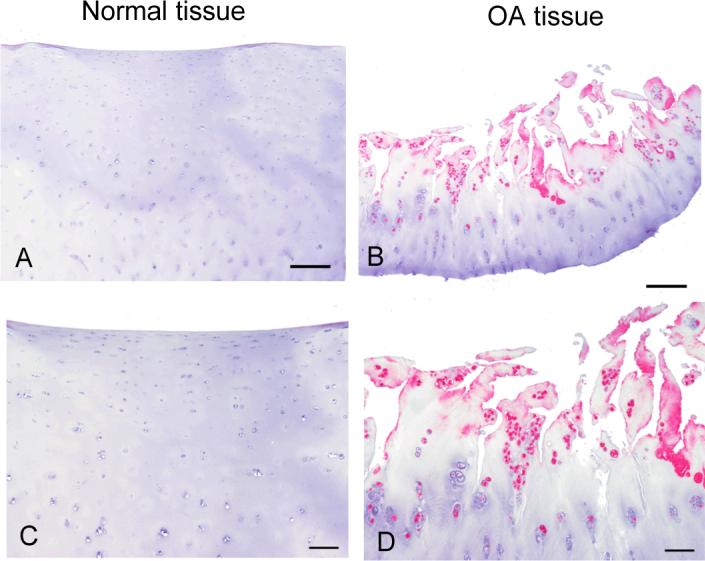
Methods: Human articular cartilage was obtained from normal tissue donors and from patients with OA at the time of knee replacement surgery. TRB3 was assessed in the tissue samples by reverse transcription-polymerase chain reaction, immunoblotting, and immunohistochemistry. Overexpression of TRB3 was induced by transient transfection to determine the effects of TRB3 on cell survival and proteoglycan synthesis.

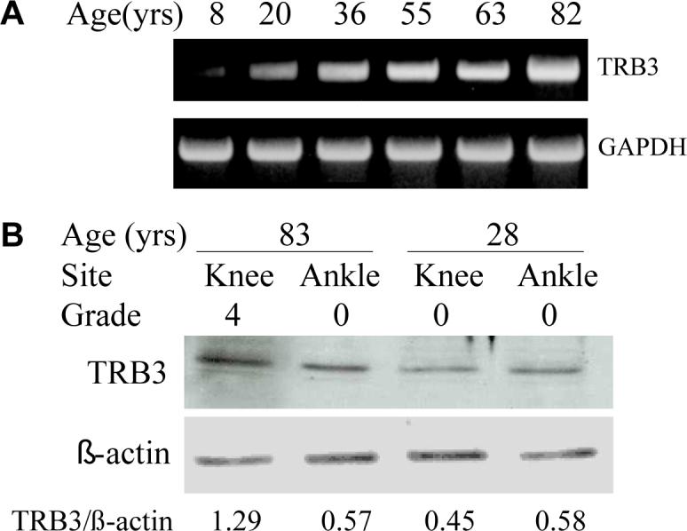
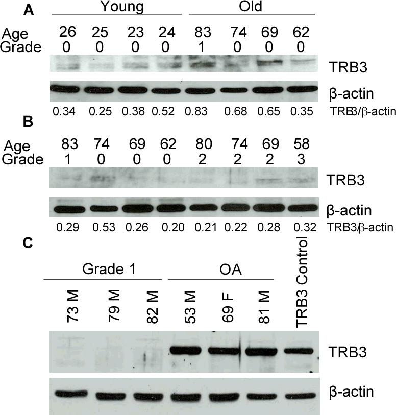
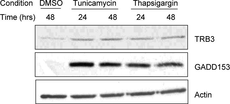
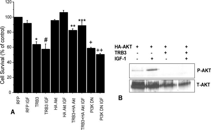
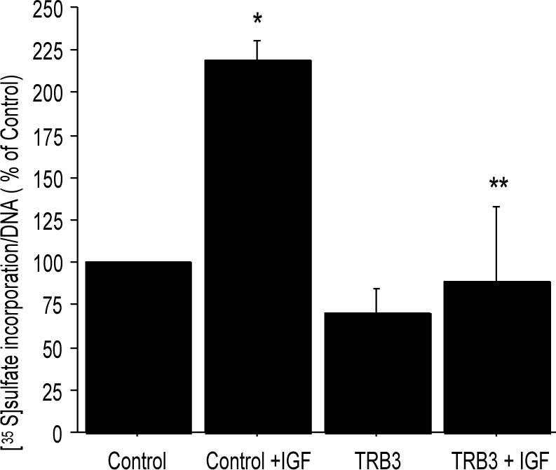
Results: TRB3 messenger RNA was detected in normal human chondrocytes. TRB3 protein levels were low in cells from normal cartilage but significantly increased in cells from OA cartilage. Incubation with 2 agents that induce endoplasmic reticulum stress, tunicamycin and thapsigargin, increased TRB3 levels in normal cells. Overexpression of TRB3 inhibited Akt phosphorylation and reduced chondrocyte survival and proteoglycan synthesis.

Conclusion: These results are the first to demonstrate that TRB3 is present in human chondrocytes, and that the level of TRB3 is increased in OA cartilage and in isolated OA chondrocytes. Because it is an inhibitor of Akt activation, elevated TRB3 production could play a role in the increased cell death and reduced response to IGF-1 observed in OA cartilage.


Figures

Figure 1
Figure 1. Chondrocytes express TRB3
(A) Human chondrocytes isolated from donors of different ages were plated in monolayer culture. At confluency, RNA was isolated from these cells, RT-PCR was performed, and the cDNA products were stained with ethidium after separation on an agarose gel. GAPDH expression was used as a control. (B) Chondrocytes were isolated from matched pairs of knee and ankle cartilage from an old (83 years) and young (28 years) donor. Cell lysates were prepared after overnight incubation in serum-free media and were used for immunoblot analysis with anti-TRB3 antibody. Immunoblotting for actin was used as a protein loading control. Densitometry was used to quantitate the band intensity which is reported beneath the blots as a ratio of TRB3 to actin.
Figure 2
Figure 2. The effect of donor age, tissue grade and OA on TRB3 protein levels
(A) Protein extracts were made directly from ankle tissue graded 0−1 and obtained from young and old tissue donors without a known history of arthritis. Immunoblot analysis was performed using anti-TRB3 antibody and with anti-β-actin antibody as a loading control. Densitometry results are shown under the blots. (B) Protein extracts prepared and analyzed as in panel A using samples obtained from older adult ankle cartilage with grades from 0−3. (C) Cell lysates were prepared from cultured, age-matched, untreated human chondrocytes from normal tissue donors and tissue harvested at time of knee joint replacement for OA. The lysates were used for immunoblot analysis with an anti-TRB3 antibody along with anti-β-actin antibody as a loading control. An additional cell lysate made from human chondrocytes transfected with a TRB3 expression vector was used as a positive control.
Figure 3
Figure 3. Immunohistochemical staining for TRB3 in normal and OA knee tissue
Immunohistochemistry was done on tissue from a normal knee joint and from one obtained at joint replacement for OA. The sections were immunostained with anti-TRB3 at a 1:500 dilution. An alkaline phosphatase detection system was used and the product was visualized with Vector Red. The negative control images (not shown) were similar to the images from normal tissue which did not detect TRB3. Bar = 250μm in the upper images and 100μm in the lower images.
Figure 4
Figure 4. ER stress induces TRB3 expression
Normal human chondrocytes cultured in monolayer were treated with ER stress inducing agents tunicamycin (2μg/mL) or thapsigargin (2μM) or 0.001% DMSO for the indicated times and cell lysates were used for immunoblot analysis with anti-TRB3 antibody, anti-GADD153 antibody (ER stress marker) and anti-β-actin antibody as a loading control.
Figure 5
Figure 5. Overexpression of TRB3 reduces chondrocyte survival and IGF-1 stimulated Akt phosphorylation
(A) Human chondrocytes isolated from articular cartilage of normal donor ankles were nucleofected with plasmid constructs to express TRB3, HA-Akt, red fluorescent protein (RFP control), or a dominant-negative PI-3 kinase. After 48 hours cells were harvested and placed into alginate beads at 20,000 cells/bead and cultured in serum-free media for 72 hours with either 50ng/ml IGF-1 or vehicle as a control. Live/Dead survival assay was then performed (n=3 experiments with cells from different donors). p <0.0001: RFP control vs TRB3 (*), RFP IGF vs TRB3 IGF (#), RFP vs PI3KDN (+), and RFP IGF to PI3KDN IGF(++). p=0.001: TRB3+HA Akt vs TRB3(**). p=0.03: TRB3+HA Akt IGF vs TRB3 IGF(***). (B) Normal human chondrocytes were transfected with an HA tagged Akt expression construct or cotransfected with HA tagged Akt and TRB3 expression constructs. After 48hours cultures were made serum free overnight. Indicated wells were treated with 100ng/mL IGF-1 for 30 minutes and then all wells were lysed. Immunoprecipitation was performed using anti-HA antibody and then the lysates were immunoblotted using anti-phosphorylated Akt antibody (P-AKT). The membrane was stripped and reprobed using anti-Akt antibody (T-AKT). This is a representative blot from an n of 4 experiments.
Figure 6
Figure 6. Overexpression of TRB3 decreases IGF-1 stimulation of PG synthesis
Normal human chondrocytes were transfected with 5μg pcDNA 3.0 empty vector or 5μg TRB3 overexpression plasmids. The cells were allowed to recover from the transfection in monolayer culture and then placed in media containing 5% serum. 100ng/mL IGF-1 was added to indicated wells for an overnight treatment and then [35S]sulfate incorporation was performed. The counts per minute were normalized to DNA concentration of the samples and the results (mean±SEM) expressed as a % of the unstimulated control (n=3 experiments with cells from different donors). *p=0.02 control vs control+IGF-1; **p=0.02 control+IGF-1 vs TRB3+IGF-1.

Similar articles

Cited by

References

    1. Aigner T, Haag J, Martin J, Buckwalter J. Osteoarthritis: aging of matrix and cells - going for a remedy. Current Drug Targets. 2007;8:325–331. - PubMed
    1. Goldring MB. The role of the chondrocyte in osteoarthritis. Arthritis Rheum. 2000;43(9):1916–26. - PubMed
    1. Loeser RF, Shanker G, Carlson CS, Gardin JF, Shelton BJ, Sonntag WE. Reduction in the chondrocyte response to insulin-like growth factor 1 in aging and osteoarthritis: studies in a non-human primate model of naturally occurring disease. Arthritis Rheum. 2000;43(9):2110–20. - PubMed
    1. van Osch GJ, van den Berg WB, Hunziker EB, Hauselmann HJ. Differential effects of IGF-1 and TGF beta-2 on the assembly of proteoglycans in pericellular and territorial matrix by cultured bovine articular chondrocytes. Osteoarthritis Cartilage. 1998;6(3):187–95. - PubMed
    1. Guenther HL, Guenther HE, Froesch ER, Fleisch H. Effect of insulin-like growth factor on collagen and glycosaminoglycan synthesis by rabbit articular chondrocytes in culture. Experientia. 1982;38(8):979–81. - PubMed

Publication types

MeSH terms