. 2009 Apr;37(6):1817-28.
doi: 10.1093/nar/gkp018. Epub 2009 Jan 30.

Mitochondrial DNA level, but not active replicase, is essential for Caenorhabditis elegans development

Affiliations

Mitochondrial DNA level, but not active replicase, is essential for Caenorhabditis elegans development

Ivana Bratic et al. Nucleic Acids Res. 2009 Apr.

Abstract

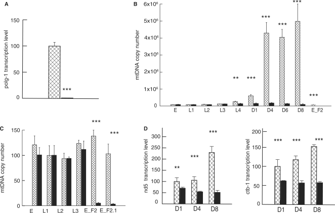
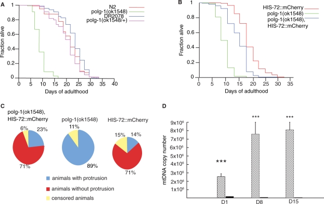
A number of studies showed that the development and the lifespan of Caenorhabditis elegans is dependent on mitochondrial function. In this study, we addressed the role of mitochondrial DNA levels and mtDNA maintenance in development of C. elegans by analyzing deletion mutants for mitochondrial polymerase gamma (polg-1(ok1548)). Surprisingly, even though previous studies in other model organisms showed necessity of polymerase gamma for embryonic development, homozygous polg-1(ok1548) mutants had normal development and reached adulthood without any morphological defects. However, polg-1 deficient animals have a seriously compromised gonadal function as a result of severe mitochondrial depletion, leading to sterility and shortened lifespan. Our results indicate that the gonad is the primary site of mtDNA replication, whilst the mtDNA of adult somatic tissues mainly stems from the developing embryo. Furthermore, we show that the mtDNA copy number shows great plasticity as it can be almost tripled as a response to the environmental stimuli. Finally, we show that the mtDNA copy number is an essential limiting factor for the worm development and therefore, a number of mechanisms set to maintain mtDNA levels exist, ensuring a normal development of C. elegans even in the absence of the mitochondrial replicase.


Figures

Figure 1.
Figure 1.
Steady-state levels of mtDNA and mitochondrial transcripts in wild-type (N2, squared bars) and polg-1(ok1548) animals (black bars). (A) Relative transcript levels of polg-1 gene presented as a percentage of the wild type measured at day 1 of adulthood. (B, C) Steady-state mtDNA levels during worm development. Values represent the mtDNA copy number per animal (B) or per nuclear genome (C). (D) Quantification of transcript levels for mitochondrial-encoded nd5 and ctb-1 genes in polg-1(ok1548) animals at day 1 (D1), day 4 (D4) and day 8 (D8) of adulthood. Values are presented in the percentage relative to the wild-type levels at adult day 1. The error bars represent the SD. Asterisks show statistical significance (Student's t-test, **P < 0.01, ***P < 0.001).
Figure 2.
Figure 2.
Mitochondrial morphology in the body wall muscle of (A) control (myo-3::MTS::GFP) and (B) polg-1 deficient (polg-1(ok1548),myo-3::MTS::GFP) adult hermaphrodite animals. (C) Distribution of different mitochondrial lengths “+” stands for polg-1(ok1548) animals and “°” stands for N2 animals.
Figure 3.
Figure 3.
Gonad morphology of wild-type (N2) and polg-1(ok1548) animals. Electron micrographs of gonads isolated from worms at first (A) and sixth (B) day of adulthood; m: mitochondria, n: nuclei. (C) 3D representation of the gonad morphology in wild-type (N2) and polg-1(ok1548) mutant adult hermaphrodites. 3D reconstruction of microscopic images was performed after Hoechst staining of isolated gonads with the software Endrov (ver. 2.11.0).
Figure 4.
Figure 4.
Embryonic development of polg-1(ok1548) progeny. (A) Quantification of the number of embryos arrested on specific stages during embryonic development. (B) Time intervals needed for embryonic development of wild-type (N2, squared bars) and polg-1(ok1548) (black bars) progeny to reach the two-cell, four-cell stage, the division of the EMS cell (div.EMS), of the P3 cell (div.P3), of the E cell (div.E) and the Ep cell (div.Ep).
Figure 5.
Figure 5.
Lifespan analysis of polg-1(ok1548) animals. (A) Lifespan of polg-1(ok1548) mutant was shown in comparison with DR2078, wild-type (N2) and polg-1(ok1548/+) animals. (B) Lifespan analysis of polg-1(ok1548), HIS-72::mCherry, with HIS-72::mCherry and polg-1(ok1548) animals used as controls. (C) Pie charts present percentage of animals that developed vulva protrusion during their lifespan. (D) mtDNA copy number in polg-1(ok1548), HIS-72::mCherry (black bars) and HIS-72::mCherry (squared bars) animals. The steady-state mtDNA levels are measured at day 1 (D1), day 8 (D8) and day 16 (D16). Values represent the mtDNA copy number per animal. The error bars represent the SD. Asterisks show statistical significance (Student's t-test, ***P < 0.001).

Similar articles

Cited by

References

    1. Kaguni LS. DNA polymerase gamma, the mitochondrial replicase. Annu. Rev. Biochem. 2004;73:293–320. - PubMed
    1. Trifunovic A, Wredenberg A, Falkenberg M, Spelbrink JN, Rovio AT, Bruder CE, Bohlooly-Y M, Gidlöf S, Oldfors A, Wibom R, et al. Premature ageing in mice expressing defective mitochondrial DNA polymerase. Nature. 2004;429:417–423. - PubMed
    1. Hudson G, Chinnery PF. Mitochondrial DNA polymerase-gamma and human disease. Hum. Mol. Genet. 2006;15:R244–R252. - PubMed
    1. Longley MJ, Graziewicz MA, Bienstock RJ, Copeland WC. Consequences of mutations in human DNA polymerase gamma. Gene. 2005;354:125–131. - PubMed
    1. Genga A, Bianchi L, Foury F. A nuclear mutant of Saccharomyces cerevisiae deficient in mitochondrial DNA replication and polymerase activity. J. Biol. Chem. 1986;261:9328–9332. - PubMed

Publication types

MeSH terms