. 2009 Feb 10;106(6):1983-8.
doi: 10.1073/pnas.0812491106. Epub 2009 Jan 30.

Tissue plasminogen activator modulates the cellular and behavioral response to cocaine

Affiliations

Tissue plasminogen activator modulates the cellular and behavioral response to cocaine

Rajani Maiya et al. Proc Natl Acad Sci U S A. .

Abstract

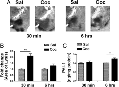
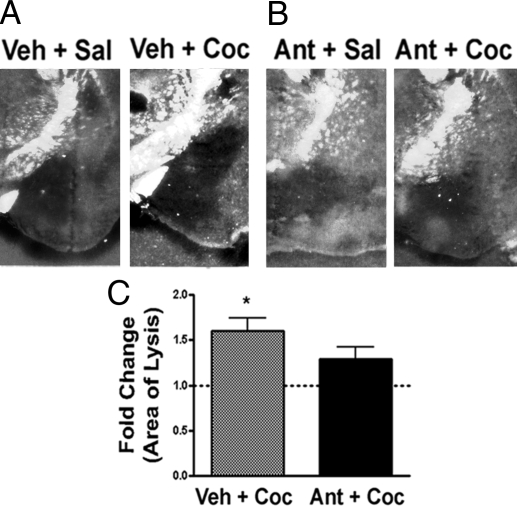
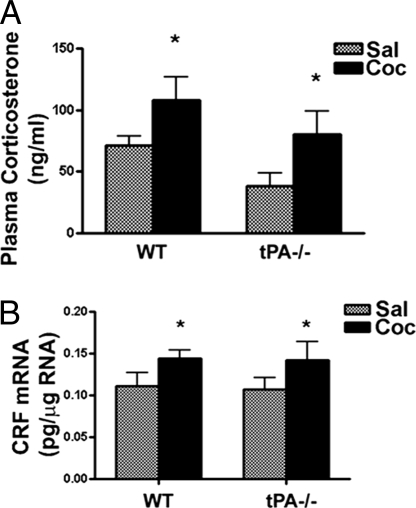
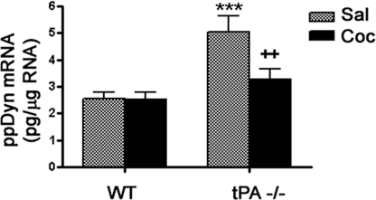
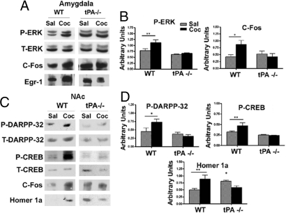
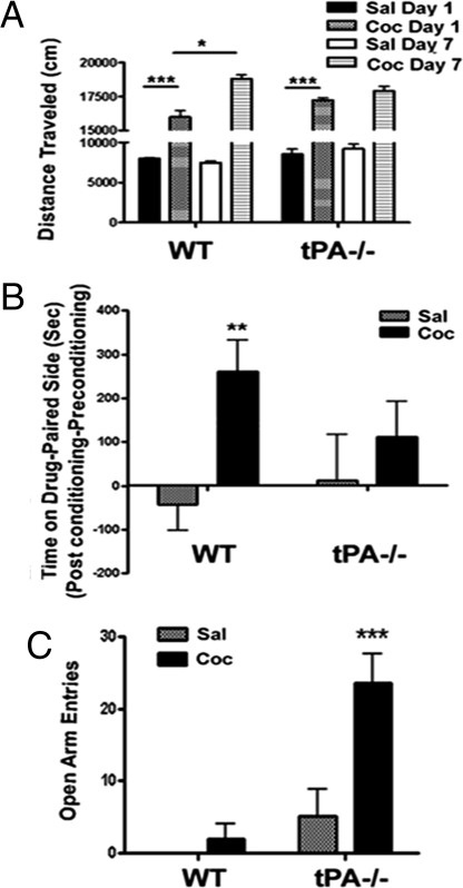
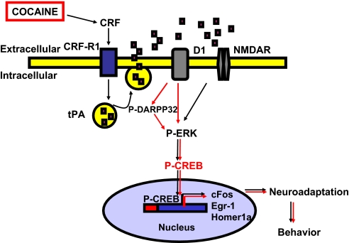
Cocaine exposure induces long-lasting molecular and structural adaptations in the brain. In this study, we show that tissue plasminogen activator (tPA), an extracellular protease involved in neuronal plasticity, modulates the biochemical and behavioral response to cocaine. When injected in the acute binge paradigm, cocaine enhanced tPA activity in the amygdala, which required activation of corticotropin-releasing factor type-1 (CRF-R1) receptors. Compared with WT mice, tPA-/- mice injected with cocaine displayed attenuated phosphorylation of ERK, cAMP response element binding protein (CREB), and dopamine and cAMP-regulated phosphoprotein 32 kDa (DARPP-32) and blunted induction of immediate early genes (IEGs) c-Fos, Egr-1, and Homer 1a in the amygdala and the nucleus accumbens (NAc). tPA-/- mice also displayed significantly higher basal preprodynorphin (ppDyn) mRNA levels in the NAc in comparison to WT mice, and cocaine decreased ppDyn mRNA levels in tPA-/- mice only. Cocaine-induced locomotor sensitization and conditioned place preference (CPP) were attenuated in tPA-/- mice. Cocaine exposure also had an anxiolytic effect in tPA-/- but not WT mice. These results identify tPA as an important and novel component of the signaling pathway that modulates cocaine-induced changes in neuroadaptation and behavior.


Conflict of interest statement

The authors declare no conflict of interest.

Figures

Fig. 1.
Fig. 1.
Acute cocaine exposure increases tPA activity in the amygdala. (A) Extracellular tPA activity was measured by in situ zymography 30 min and 6 h after the final cocaine injection and quantified by measuring area of lysis (dark lytic zones). tPA activity was significantly increased in the amygdala of WT mice 30 min but not 6 h after acute binge cocaine exposure compared to saline-injected mice. (B) Quantification of results in A. Two-way ANOVA analysis revealed a significant main effect of time [F (1, 12) = 9.01, P < 0.05], treatment [F (1, 12) = 25.53, P < 0.005], and treatment × time interaction [F (1, 12) = 9.01, P < 0.05]; **,P < 0.005, cocaine vs. saline, n = 4–5/group. (C) Acute cocaine exposure increased PAI-1 levels in the amygdala 6 h but not 30 min after the last cocaine injection compared to saline-injected control samples. **, P < 0.005, t test, vs. saline control, n = 4–5/group. Sal, saline; Coc, cocaine.
Fig. 2.
Fig. 2.
Acute cocaine increases tPA activity in the amygdala via a CRF-dependent mechanism. Extracellular tPA activity was measured by in situ zymography 30 min after saline or cocaine administration. (A) tPA activity increased 1.6-fold in the amygdala of WT mice injected with vehicle plus cocaine in comparison to mice injected with vehicle plus saline. (B) This increase was prevented when mice were injected with antalarmin plus cocaine. (C) Quantification of areas of lysis in A and B. The dashed line represents tPA activity in saline-injected control mice. *, P < 0.05 compared to saline control, t test, n = 5–6/group. Sal, saline; Coc, cocaine; Veh, vehicle; Ant, antalarmin.
Fig. 3.
Fig. 3.
Acute binge cocaine increased plasma CORT and CRF mRNA levels in the amygdala of WT and tPA−/− mice. Plasma CORT levels and CRF mRNA levels were measured 30 min after the final cocaine injection. (A) Plasma CORT levels increased after acute binge cocaine administration in both genotypes. Two-way ANOVA showed a significant effect for drug treatment [F (1, 29) = 5.61, P < 0.05]; n = 8–9/group. (B) Acute binge cocaine increased CRF mRNA levels. Two-way ANOVA showed a significant effect for drug treatment [F (1, 20) = 4.44, P < 0.05]; n = 6/group. Sal, saline; Coc, cocaine.
Fig. 4.
Fig. 4.
Cocaine regulation of ppDyn levels in the NAc of WT and tPA−/− mice. ppDyn mRNA levels were measured 30 min after the final cocaine injection. Two-way ANOVA showed significant effects for drug treatment [F (1, 20) = 5.00, P < 0.05)], genotype [F (1, 20) = 16.6, P < 0.001], and drug treatment × genotype interaction [F (1, 20) = 4.62, P < 0.05]. ppDyn mRNA levels were significantly increased in the NAc of tPA -/- mice after saline injection compared to that of WT mice (P < 0.0001). After cocaine administration, ppDyn mRNA levels were significantly reduced in the NAc of tPA−/− mice in comparison to saline-injected mice (P < 0.01). ***, P < 0.001 between WT and tPA−/− mice after saline treatment; ++, P < 0.01 between cocaine vs. saline treatment in tPA−/− mice, n = 6/group. Sal, saline; Coc, cocaine.
Fig. 5.
Fig. 5.
tPA mediates the cellular response to cocaine in the amygdala and NAc. (A) Representative blots for cocaine-induced changes in neuronal signaling in the amygdala. (B) Quantification of results in A. Two-way ANOVA showed significant effects for drug treatment [F (1, 16) = 7.71, P < 0.05], genotype [F (1, 16) = 18.92, P < 0.001], and drug treatment × genotype interaction [F (1, 16) = 4.54, P < 0.05] for ERK phosphorylation after binge cocaine. Neumann–Keuls post hoc analysis indicated significant increase in P-ERK in the amygdala after binge cocaine (P < 0.005). Two-way ANOVA revealed significant interaction between genotype and drug treatment for c-Fos induction in the amygdala after acute cocaine [F (1, 13) = 5.71, P < 0.05]. Planned comparison test revealed significant increase in c-Fos induction in WT mice only after binge cocaine (P < 0.05), n = 4–6/group. Quantification of Egr-1 is presented in Fig. S3. (C) Representative blots of cocaine-induced changes in neuronal signaling in the NAc. (D) Quantification of results in C. Two-way ANOVA revealed a significant main effect of genotype [F (1, 19) = 10.88, P < 0.005] and significant genotype × treatment interaction for P-DARPP-32 levels [F (1, 19) = 5.15, P < 0.05] in the NAc. Significant increase in P-DARPP-32 after cocaine treatment was observed in WT mice only (P < 0.05). Two-way ANOVA revealed significant effect of genotype [F (1, 14) = 21.1, P < 0.0005] and genotype × treatment interaction [F (1, 14) = 5.84, P < 0.05] for cocaine induction of P-CREB levels. Cocaine enhanced P-CREB significantly in the NAc of WT mice only (P < 0.05). Two-way ANOVA also revealed a significant genotype × treatment interaction [F (1, 13) = 15.36, P < 0.005] for cocaine-induced increase in Homer 1a in the NAc. Post hoc analysis revealed significant increases in Homer 1a in cocaine-injected WT but not tPA−/− mice (P < 0.05) and in its basal expression in tPA−/− mice compared to WT (P < 0.05), n = 4–6 animals/group. Sal, saline; Coc, cocaine. Quantification of c-Fos is presented in Fig. S3.
Fig. 6.
Fig. 6.
tPA modulates the behavioral response to cocaine. (A) Distance traveled on days 1 and 7 immediately after the last saline or cocaine injection is shown for both genotypes. Acute cocaine exposure enhanced locomotor activity to a similar extent in WT and tPA−/− mice compared to saline-injected groups. ***, P < 0.0001 compared to saline control, n = 6/group. However, cocaine induced sensitization in WT (WT cocaine days 1 vs. 7) (*, P < 0.05, t test) but not tPA −/− mice. (B) Cocaine-injected WT mice spent significantly more time in the drug-paired environment in comparison to their saline-injected counterparts. **, P < 0.005, compared to WT saline, n = 7–8/group. Cocaine-injected tPA−/− mice, however, did not show significant preference to the drug-paired environment compared to their saline-injected counterparts (P = 0.2 compared to tPA−/− saline, n = 8–9/group). (C) Anxiety levels were measured using the elevated plus maze. Cocaine-injected tPA−/− mice made significantly more OAEs compared to saline-injected tPA−/− mice. ***, P < 0.005, n = 6. Saline-injected WT mice did not make any OAEs. Significant differences were not observed in the number of OAEs made by saline-treated WT and tPA−/− mice (P = 0.2). Significant differences were not observed in the number of closed arm entries between WT and tPA−/− mice after acute cocaine exposure (data not shown). Sal, saline; Coc, cocaine.
Fig. 7.
Fig. 7.
Schematic representation of tPA's role in acute cocaine-induced neuronal signaling. Signaling events modulated by tPA in the amygdala are indicated by black arrows, and those that occur in the NAc are indicated by red arrows. Components of the signaling pathway hypothesized to be altered in tPA−/− mice are shaded in gray. In the amygdala, acute cocaine exposure increases extracellular tPA activity in a CRF-dependent manner. tPA released into the extracellular space is hypothesized to modulate DA D1 receptor-mediated signaling to activate IEGs. In the NAc, tPA may modulate signaling through both D1 and NMDAR to modulate IEG expression. In the absence of tPA, cocaine-induced IEG expression is attenuated, thereby suggesting that tPA is an important component of the signaling cascade modulating cocaine-induced neuroadaptation.

Similar articles

Cited by

References

    1. Nestler EJ. Historical review: molecular and cellular mechanisms of opiate and cocaine addiction. Trends Pharmacol Sci. 2004;25(4):210–218. - PubMed
    1. Kreek MJ, LaForge KS, Butelman E. Pharmacotherapy of addictions. Nat Rev Drug Discov. 2002;1(9):710–726. - PubMed
    1. Koob G, Kreek MJ. Stress, dysregulation of drug reward pathways, and the transition to drug dependence. Am J Psychiatry. 2007;164(8):1149–1159. - PMC - PubMed
    1. Shippenberg TS, Zapata A, Chefer VI. Dynorphin and the pathophysiology of drug addiction. Pharmacol Ther. 2007;116(2):306–321. - PMC - PubMed
    1. Spangler R, Unterwald EM, Kreek MJ. ‘Binge’ cocaine administration induces a sustained increase of prodynorphin mRNA in rat caudate-putamen. Brain Res Mol Brain Res. 1993;19(4):323–327. - PubMed

Publication types

Substances

LinkOut - more resources