. 2009 Apr;8(4):595-605.
doi: 10.1128/EC.00352-08. Epub 2009 Jan 30.

Isolation and characterization of Cryptococcus neoformans spores reveal a critical role for capsule biosynthesis genes in spore biogenesis

Affiliations

Isolation and characterization of Cryptococcus neoformans spores reveal a critical role for capsule biosynthesis genes in spore biogenesis

Michael R Botts et al. Eukaryot Cell. 2009 Apr.

Abstract

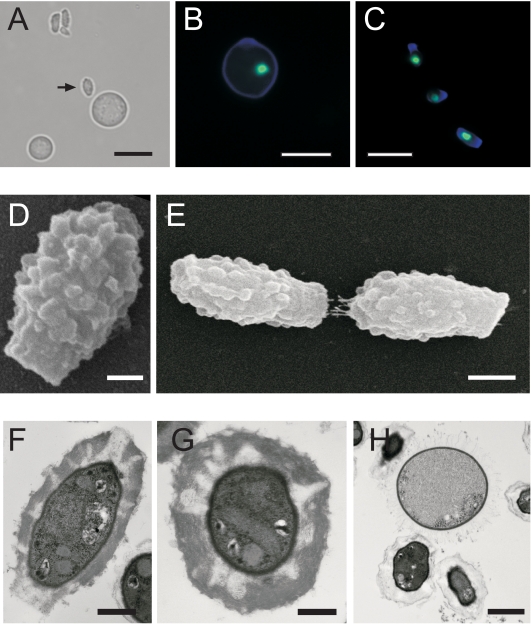
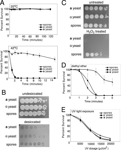
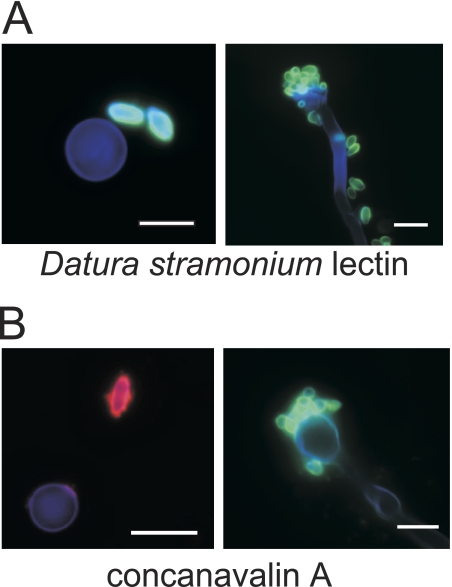
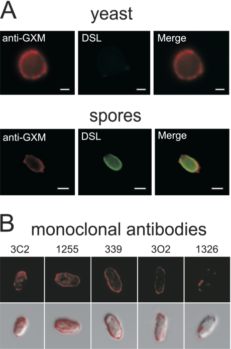
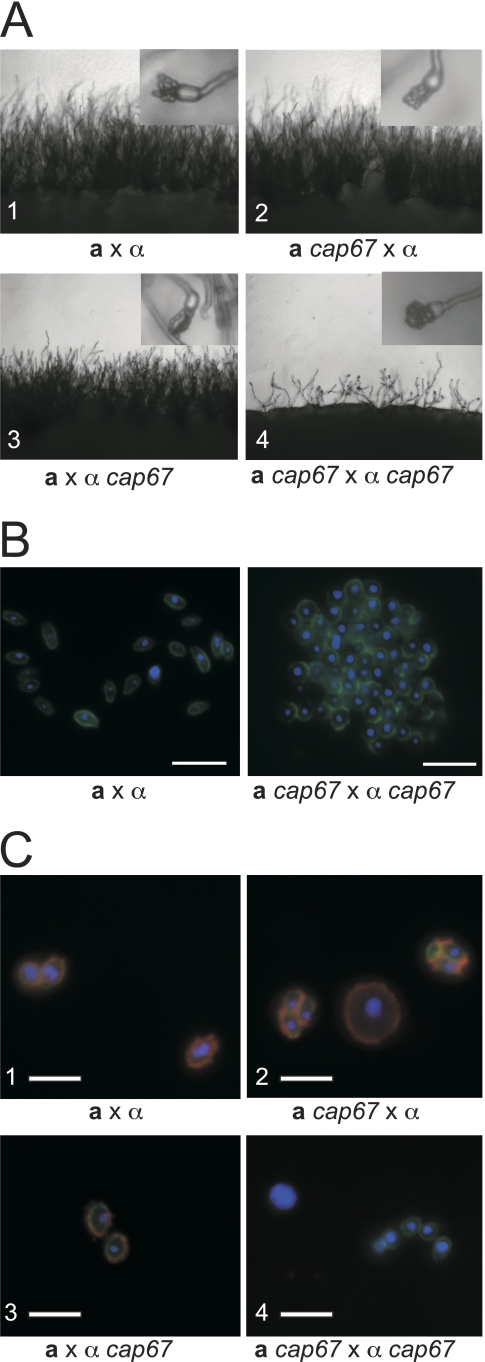
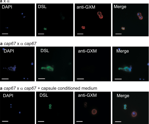
Spores are essential particles for the survival of many organisms, both prokaryotic and eukaryotic. Among the eukaryotes, fungi have developed spores with superior resistance and dispersal properties. For the human fungal pathogens, however, relatively little is known about the role that spores play in dispersal and infection. Here we present the purification and characterization of spores from the environmental fungus Cryptococcus neoformans. For the first time, we purified spores to homogeneity and assessed their morphological, stress resistance, and surface properties. We found that spores are morphologically distinct from yeast cells and are covered with a thick spore coat. Spores are also more resistant to environmental stresses than yeast cells and display a spore-specific configuration of polysaccharides on their surfaces. Surprisingly, we found that the surface of the spore reacts with antibodies to the polysaccharide glucuronoxylomannan, the most abundant component of the polysaccharide capsule required for C. neoformans virulence. We explored the role of capsule polysaccharide in spore development by assessing spore formation in a series of acapsular strains and determined that capsule biosynthesis genes are required for proper sexual development and normal spore formation. Our findings suggest that C. neoformans spores may have an adapted cell surface that facilitates persistence in harsh environments and ultimately allows them to infect mammalian hosts.


Figures

FIG. 1.
FIG. 1.
C. neoformans spores have a morphology distinct from that of yeast cells. (A) Bright-field micrograph revealing the relative differences in size and shape between a spore (arrow) and a nearby yeast cell. Bar, 5 μm. (B and C) Yeast cell (B) and spores (C) stained with calcofluor white (blue cell walls) and SYTOX green (green nuclei). Bars, 5 μm. (D) SEM micrograph showing the ultrastructure of the spore. Bar, 500 nm.(E) SEM micrograph showing two spores end to end. Bar, 1 μm. (F) TEM micrograph of a longitudinal section of a spore. Bar, 500 nm. (G) TEM micrograph of a latitudinal section of a spore. Bar, 250 nm. (H) TEM micrograph of two spores adjacent to a yeast cell. Bar, 1 μm.
FIG. 2.
FIG. 2.
Spores are more resistant to environmental stress than yeast cells. All environmental stress resistance experiments were carried out using wild-type a and α yeast cells (JEC20 and JEC21, respectively) and spores purified from a cross between them. (A) Resistance to heat shock. Both a and α yeast cells (circles and squares, respectively) and spores (triangles) were incubated in PBS at 25°C or 42°C before they were plated on rich growth medium. On each graph, the x axis shows time of incubation and the y axis shows percent survival. (B) Resistance to desiccation. The top panel shows 10-fold serial dilutions of spores and parental yeast cells grown on rich medium. The bottom panel shows the same dilutions spotted onto a nitrocellulose membrane and incubated at room temperature for 2 days before the membrane was transferred to rich medium. Rows are labeled according to cell type (top, a yeast; middle, α yeast; and bottom, spores). (C) Resistance to oxidative stress. Both a and α yeast cells and spores were diluted in 10-fold serial dilutions and incubated in 10 mM H2O2 before they were plated to rich medium. Rows are labeled according to cell type (top, a yeast; middle, α yeast; and bottom, spores). (D) Resistance to diethyl ether. Both a and α yeast cells and spores were plated on rich growth medium and then exposed to diethyl ether vapors. The x axis shows time of exposure, and the y axis shows percent survival of spores and yeast cells after vapor exposure. (E) Exposure to UV light. Both a and α yeast cells and spores were diluted to 300 cells in 10 μl water and spotted on a solid surface. The spots were exposed to increasing dosages of UV light before they were plated to rich medium. The x axis shows the dose of UV in μJ/cm2, and the y axis shows percent survival compared to untreated cells.
FIG. 3.
FIG. 3.
Spores have specific lectin binding properties. Spores and yeast cells were stained with calcofluor white (to stain all cell types) and fluorescently labeled lectins (to show spore-specific binding). Stained cells were visualized by fluorescence microscopy. Bars, 5 μm. (A) Spore-specific staining by DSL (green), which binds N-acetylglucosamine oligomers. The left panel shows a yeast cell (∼5 μm in diameter; blue) and two spores (∼3 μm long; green). The right panel shows a long filament cell with a terminal basidium (club-like structure of ∼5 μm in diameter; blue), with spores from collapsed chains on the end and scattered along the filament (∼3 μm long; green). (B) Spore-specific staining by ConA (visualized in red in the left panel in and green in the right panel), which binds α-mannose residues. The left panel shows a yeast cell (∼5 μm in diameter; blue) and a spore (∼3 μm long; red). The right panel shows a basidium (club-like structure of ∼5 μm in diameter; blue) with collapsed spore chains attached (∼3 μm long; green).
FIG. 4.
FIG. 4.
The spore surface is bound by antibodies to the capsular polysaccharide GXM. (A) The top panels show a yeast cell stained with the anti-GXM antibody F12D2 (red) and with DSL (no signal). The bottom panels show an identically stained spore brightly labeled with both anti-GXM antibody (red) and DSL (green), indicating the presence of anti-GXM epitopes on the surfaces of spores. Bars, 1 μm. (B) The top panels show the binding of five independent monoclonal anti-GXM antibodies (red). The bottom panels display a merge of anti-GXM antibody staining with differential interference contrast bright-field images of spores.
FIG. 5.
FIG. 5.
Capsule mutants have defects in sexual development and spore formation. (A) All panels show the periphery of a cross on V8 medium under a light microscope at low magnification (×200). Insets each show a single basidium under higher magnification (×400). (1) Wild-type a × α cross. (2 and 3) Wild-type a or α strain crossed with corresponding a cap67 or α cap67 strain. (4) a cap67 × α cap67 cross. The few basidia that were produced did not develop proper spore chains and yielded clumps of spores (inset in panel 4). Identical results were obtained for all additional capsule deletion strains tested (cap10Δ, cap60Δ, and cap64Δ strains). (B) Spores were stained with DAPI (blue) and DSL (green) and visualized by fluorescence micros- copy. (Left) Normal morphology of spores derived from a wild-type cross. (Right) Spores derived from a cap67 × cap67 cross forming an aggregated mass of spores. (C) Spores from wild-type and cap67 strains stained with DAPI to reveal nuclei (blue), with anti-GXM antibody (F12D2) to detect capsule (red), and with DSL to identify spores (green) were visualized by immunofluorescence microscopy. All three color channels were merged into a single image for each sample. (1) Spores derived from a wild-type a by α cross (JEC20 × JEC21). (2 and 3) Spores and yeast cells from crosses between wild-type and cap67 strains (CHY1377 × JEC21 and JEC20 × ATCC 52817, respectively). (4) Spores and yeast cells isolated from a cross between two cap67 strains (CHY1377 × ATCC 52817). Bars, 5 μm.
FIG. 6.
FIG. 6.
Capsule is not efficiently transferred to the surfaces of capsule-deficient spores. Yeast cells and spores were labeled with DAPI (blue) to reveal nuclei, with DSL (green) to identify spores, and with anti-GXM antibody F12D2 (red) to highlight polysaccharide epitopes. The top row of panels shows yeast cells and spores from a wild-type cross. Note that wild-type spores and yeast cells have similar intensities of staining with anti-GXM antibody. The middle row shows yeast cells and spores from an a cap67 × α cap67 cross, in which no anti-GXM antibody staining is detectable. The bottom row of panels shows yeast cells and spores stained after incubation in capsule-conditioned medium. Anti-GXM antibody staining shows brightly stained yeast cells, with only faint staining of the spores. The final panel in each row is a merged image of all three fluorescence channels used.

Similar articles

Cited by

References

    1. Alspaugh, J. A., J. R. Perfect, and J. Heitman. 1998. Signal transduction pathways regulating differentiation and pathogenicity of Cryptococcus neoformans. Fungal Genet. Biol. 251-14. - PubMed
    1. Arrage, A. A., T. J. Phelps, R. E. Benoit, and D. C. White. 1993. Survival of subsurface microorganisms exposed to UV radiation and hydrogen peroxide. Appl. Environ. Microbiol. 593545-3550. - PMC - PubMed
    1. Brandt, S., P. Thorkildson, and T. R. Kozel. 2003. Monoclonal antibodies reactive with immunorecessive epitopes of glucuronoxylomannan, the major capsular polysaccharide of Cryptococcus neoformans. Clin. Diagn. Lab. Immunol. 10903-909. - PMC - PubMed
    1. Briza, P., M. Breitenbach, A. Ellinger, and J. Segall. 1990. Isolation of two developmentally regulated genes involved in spore wall maturation in Saccharomyces cerevisiae. Genes Dev. 41775-1789. - PubMed
    1. Briza, P., A. Ellinger, G. Winkler, and M. Breitenbach. 1988. Chemical composition of the yeast ascospore wall. The second outer layer consists of chitosan. J. Biol. Chem. 26311569-11574. - PubMed

Publication types

MeSH terms

LinkOut - more resources