

Motion of the shoulder complex during multiplanar humeral elevation
- PMID: 19181982
- PMCID: PMC2657311
- DOI: 10.2106/JBJS.G.01483
Motion of the shoulder complex during multiplanar humeral elevation
Abstract
Background: Many prior studies have evaluated shoulder motion, yet no three-dimensional analysis comparing the combined clavicular, scapular, and humeral motion during arm elevation has been done. We aimed to describe and compare dynamic three-dimensional motion of the shoulder complex during raising and lowering the arm across three distinct elevation planes (flexion, scapular plane abduction, and coronal plane abduction).
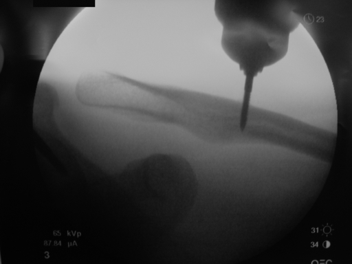
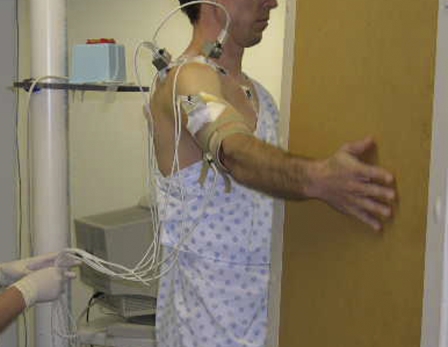
Methods: Twelve subjects without a shoulder abnormality were enrolled. Transcortical pin placement into the clavicle, scapula, and humerus allowed electromagnetic motion sensors to be rigidly fixed. The subjects completed two repetitions of raising and lowering the arm in flexion, scapular, and abduction planes. Three-dimensional angles were calculated for sternoclavicular, acromioclavicular, scapulothoracic, and glenohumeral joint motions. Joint angles between humeral elevation planes and between raising and lowering of the arm were compared.
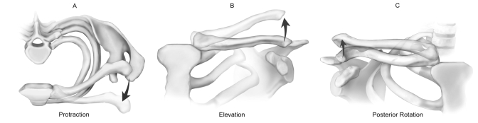
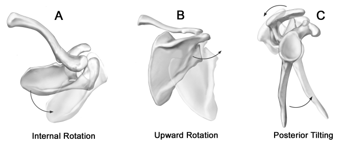
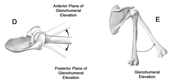
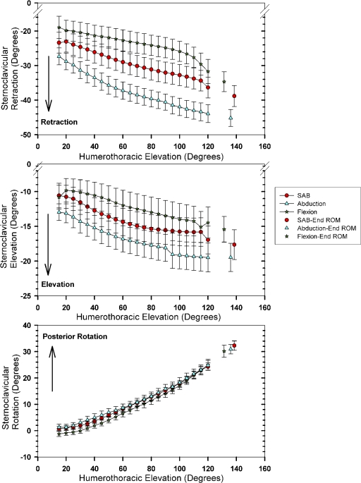
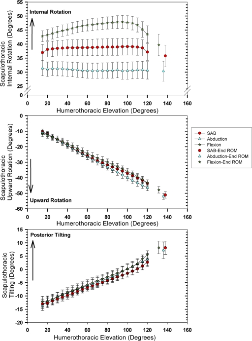
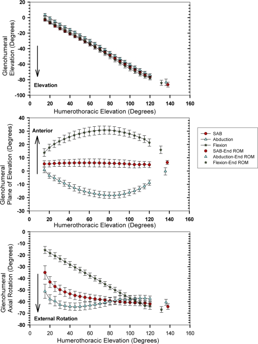
Results: General patterns of shoulder motion observed during humeral elevation were clavicular elevation, retraction, and posterior axial rotation; scapular internal rotation, upward rotation, and posterior tilting relative to the clavicle; and glenohumeral elevation and external rotation. Clavicular posterior rotation predominated at the sternoclavicular joint (average, 31 degrees). Scapular posterior tilting predominated at the acromioclavicular joint (average, 19 degrees). Differences between flexion and abduction planes of humerothoracic elevation were largest for the glenohumeral joint plane of elevation (average, 46 degrees).
Conclusions: Overall shoulder motion consists of substantial angular rotations at each of the four shoulder joints, enabling the multiple-joint interaction required to elevate the arm overhead.
Figures

Similar articles
-
Comparison of 3-dimensional shoulder complex kinematics in individuals with and without shoulder pain, part 1: sternoclavicular, acromioclavicular, and scapulothoracic joints.J Orthop Sports Phys Ther. 2014 Sep;44(9):636-45, A1-8. doi: 10.2519/jospt.2014.5339. Epub 2014 Aug 7. J Orthop Sports Phys Ther. 2014. PMID: 25103135 Free PMC article.
-
Three-dimensional clavicular motion during arm elevation: reliability and descriptive data.J Orthop Sports Phys Ther. 2004 Mar;34(3):140-9. doi: 10.2519/jospt.2004.34.3.140. J Orthop Sports Phys Ther. 2004. PMID: 15089027
-
The Coupled Kinematics of Scapulothoracic Upward Rotation.Phys Ther. 2020 Feb 7;100(2):283-294. doi: 10.1093/ptj/pzz165. Phys Ther. 2020. PMID: 31696926 Free PMC article.
-
Shoulder muscle activity and function in common shoulder rehabilitation exercises.Sports Med. 2009;39(8):663-85. doi: 10.2165/00007256-200939080-00004. Sports Med. 2009. PMID: 19769415 Review.
-
The shoulder joint complex in the throwing motion.J Shoulder Elbow Surg. 2024 Feb;33(2):443-449. doi: 10.1016/j.jse.2023.06.031. Epub 2023 Jul 26. J Shoulder Elbow Surg. 2024. PMID: 37499784 Review.
Cited by
-
[Instability pattern of acromioclavicular joint dislocations type Rockwood III: relevance of horizontal instability].Orthopade. 2013 Apr;42(4):271-7. doi: 10.1007/s00132-013-2085-1. Orthopade. 2013. PMID: 23512005 Clinical Trial. German.
-
Relationship Between Skin Intrinsic Fluorescence--an Indicator of Advanced Glycation End Products-and Upper Extremity Impairments in Individuals With Diabetes Mellitus.Phys Ther. 2015 Aug;95(8):1111-9. doi: 10.2522/ptj.20140340. Epub 2015 Apr 9. Phys Ther. 2015. PMID: 25858973 Free PMC article.
-
Comparing trapezius muscle activity in the different planes of shoulder elevation.J Phys Ther Sci. 2015 May;27(5):1495-7. doi: 10.1589/jpts.27.1495. Epub 2015 May 26. J Phys Ther Sci. 2015. PMID: 26157248 Free PMC article.
-
Effect of trapezius muscle strength on three-dimensional scapular kinematics.J Phys Ther Sci. 2016 Jun;28(6):1864-7. doi: 10.1589/jpts.28.1864. Epub 2016 Jun 28. J Phys Ther Sci. 2016. PMID: 27390435 Free PMC article.
-
Band Pull-Apart Exercise: Effects of Movement Direction and Hand Position on Shoulder Muscle Activity.Int J Sports Phys Ther. 2022 Apr 2;17(3):400-408. doi: 10.26603/001c.33026. eCollection 2022. Int J Sports Phys Ther. 2022. PMID: 35391860 Free PMC article.
References
-
- Inman VT, Saunders JB, Abbott LC. Observations on the function of the shoulder joint. J Bone Joint Surg Am. 1944;26:1-30.
-
- Dvir Z, Berme N. The shoulder complex in elevation of the arm: a mechanism approach. J Biomech. 1978;11:219-25. - PubMed
-
- Freedman L, Munro RR. Abduction of the arm in the scapular plane: scapular and glenohumeral movements. J Bone Joint Surg Am. 1966;48:1503-10. - PubMed
-
- Poppen NK, Walker PS. Normal and abnormal motion of the shoulder. J Bone Joint Surg Am. 1976;58:195-201. - PubMed
-
- Ludewig PM, Cook TM, Nawoczenski DA. Three-dimensional scapular orientation and muscle activity at selected positions of humeral elevation. J Orthop Sports Phys Ther. 1996;24:57-65. - PubMed
Publication types
MeSH terms
Grants and funding
LinkOut - more resources
Full Text Sources
Other Literature Sources
Molecular Biology Databases
